
Page 175 - Clinical Anatomy
P. 175
ECA3 7/18/06 6:44 PM Page 160
160 The upper limb
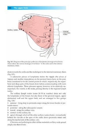
Fig. 113◊Diagram of the principal pathways of lymphatic drainage of the breast.
These follow the venous drainage of the breast—to the axilla and to the internal
mammary chain.
drain towards the axilla and the medial part to the internal mammary chain
(Fig. 113).
A subareolar plexus of lymphatics below the nipple (the plexus of
Sappey) and another deep plexus on the pectoral fascia have, in the past,
been considered to be the central points to which, respectively, the super-
ficial and deep parts of the breast drain before communicating with main
efferent lymphatics. These plexuses appear, however, to be relatively un-
important, the vessels, in the main, passing directly to the regional lymph
nodes.
The axillary lymph nodes (some 20–30 | in number) drain not only
the lymphatics of the breast, but also those of the pectoral region, upper
abdominal wall and the upper limb, and are arranged in five groups
(Fig. 114):
1◊◊anterior—lying deep to pectoralis major along the lower border of pec-
toralis minor;
2◊◊posterior—along the subscapular vessels;
3◊◊lateral—along the axillary vein;
4◊◊central—in the axillary fat;
5◊◊apical (through which all the other axillary nodes drain)—immediately
behind the clavicle at the apex of the axilla above pectoralis minor and
along the medial side of the axillary vein.
Clinicians and pathologists often define metastatic axillary node spread
simply into three levels: