
Page 178 - Clinical Anatomy
P. 178
ECA3 7/18/06 6:44 PM Page 163
Surface anatomy and surface markings 163
Bones and joints (See Figs 120, 122, 123, 125)
The subcutaneous border of the clavicle can be palpated throughout
its length; the supraclavicular nerves crossing it can be rolled against the
bone.
The acromion process forms a sharp bony edge at the lateral extremity of
the scapular spine. It lies immediately above the smooth bulge of the deltoid
muscle which itself covers the greater tubercle of the humerus. Less easily iden-
tified is the coracoid process of the scapula, lying immediately below the
clavicle at the junction of the middle and outer thirds, and covered by the
anterior fibres of the deltoid.
The medial border of the scapula can be both seen and felt. Abduction of
the arm is a complex affair made up of abduction at the shoulder joint,
depression at the sternoclavicular joint and rotation of the scapula; the last
two are readily confirmed on self-palpation.
With the shoulder abducted, the head of the humerus can be felt in the
axilla; note its movement with rotation of the arm.
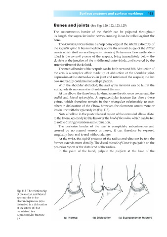
At the elbow, the three bony landmarks are the olecranon process and the
medial and lateral epicondyles. A supracondylar fracture lies above these
points, which therefore remain in their triangular relationship to each
other; in dislocation of the elbow, however, the olecranon comes more or
less in line with the epicondyles (Fig. 115).
Note a hollow in the posterolateral aspect of the extended elbow distal
to the lateral epicondyle; this lies over the head of the radius which can be felt
to rotate during pronation and supination.
The posterior border of the ulna is completely subcutaneous and
crossed by no named vessels or nerve; it can therefore be exposed
surgically from end to end without danger.
At the wrist, the styloid processes of the radius and ulna can be felt; the
former extends more distally. The dorsal tubercle of Lister is palpable on the
posterior aspect of the distal end of the radius.
In the palm of the hand, palpate the pisiform at the base of the
Fig. 115◊The relationship
of the medial and lateral
epicondyles to the
olecranon process (a) is
disturbed in a dislocation
of the elbow (b) but
maintained in a
supracondylar fracture
(c).