
Page 224 - Clinical Anatomy
P. 224
ECA4 7/18/06 6:47 PM Page 209
The anatomy and surface markings of the lower limb 209
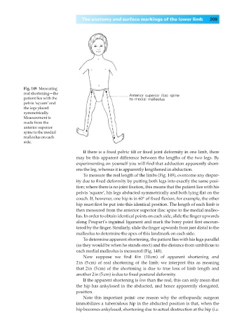
Fig. 149◊Measuring
real shortening—the
patient lies with the
pelvis ‘square’ and
the legs placed
symmetrically.
Measurement is
made from the
anterior superior
spine to the medial
malleolus on each
side.
If there is a fixed pelvic tilt or fixed joint deformity in one limb, there
may be this apparent difference between the lengths of the two legs. By
experimenting on yourself you will find that adduction apparently short-
ens the leg, whereas it is apparently lengthened in abduction.
To measure the real length of the limbs (Fig. 149), overcome any dispar-
ity due to fixed deformity by putting both legs into exactly the same posi-
tion; where there is no joint fixation, this means that the patient lies with his
pelvis ‘square’, his legs abducted symmetrically and both lying flat on the
couch. If, however, one hip is in 60° of fixed flexion, for example, the other
hip must first be put into this identical position. The length of each limb is
then measured from the anterior superior iliac spine to the medial malleo-
lus. In order to obtain identical points on each side, slide the finger upwards
along Poupart’s inguinal ligament and mark the bony point first encoun-
tered by the finger. Similarly, slide the finger upwards from just distal to the
malleolus to determine the apex of this landmark on each side.
To determine apparent shortening, the patient lies with his legs parallel
(as they would be when he stands erect) and the distance from umbilicus to
each medial malleolus is measured (Fig. 148).
Now suppose we find 4in (10cm) of apparent shortening and
2in (5cm) of real shortening of the limb; we interpret this as meaning
that 2in (5cm) of the shortening is due to true loss of limb length and
another 2in (5cm) is due to fixed postural deformity.
If the apparent shortening is less than the real, this can only mean that
the hip has ankylosed in the abducted, and hence apparently elongated,
position.
Note this important point: one reason why the orthopaedic surgeon
immobilizes a tuberculous hip in the abducted position is that, when the
hip becomes ankylosed, shortening due to actual destruction at the hip (i.e.