
Page 225 - Clinical Anatomy
P. 225
ECA4 7/18/06 6:47 PM Page 210
210 The lower limb
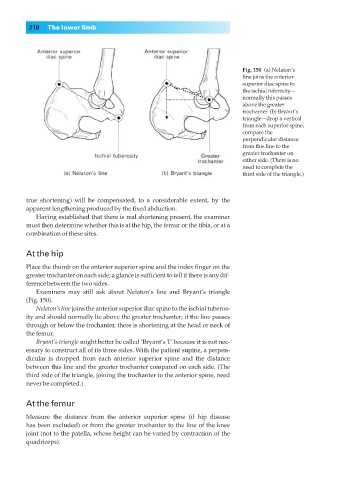
Fig. 150◊(a) Nelaton’s
line joins the anterior
superior iliac spine to
the ischial tuberosity—
normally this passes
above the greater
trochanter. (b) Bryant’s
triangle—drop a vertical
from each superior spine;
compare the
perpendicular distance
from this line to the
greater trochanter on
either side. (There is no
need to complete the
third side of the triangle.)
true shortening) will be compensated, to a considerable extent, by the
apparent lengthening produced by the fixed abduction.
Having established that there is real shortening present, the examiner
must then determine whether this is at the hip, the femur or the tibia, or at a
combination of these sites.
At the hip
Place the thumb on the anterior superior spine and the index finger on the
greater trochanter on each side; a glance is sufficient to tell if there is any dif-
ference between the two sides.
Examiners may still ask about Nelaton’s line and Bryant’s triangle
(Fig. 150).
Nelaton’s line joins the anterior superior iliac spine to the ischial tuberos-
ity and should normally lie above the greater trochanter; if the line passes
through or below the trochanter, there is shortening at the head or neck of
the femur.
Bryant’s triangle might better be called ‘Bryant’s T’ because it is not nec-
essary to construct all of its three sides. With the patient supine, a perpen-
dicular is dropped from each anterior superior spine and the distance
between this line and the greater trochanter compared on each side. (The
third side of the triangle, joining the trochanter to the anterior spine, need
never be completed.)
At the femur
Measure the distance from the anterior superior spine (if hip disease
has been excluded) or from the greater trochanter to the line of the knee
joint (not to the patella, whose height can be varied by contraction of the
quadriceps).