
Page 229 - Clinical Anatomy
P. 229
ECA4 7/18/06 6:47 PM Page 214
214 The lower limb
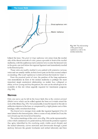
Fig. 154◊The relationship
of the great (long)
saphenous vein to the
medial malleolus.
behind the knee. The great (or long) saphenous vein arises from the medial
side of the dorsal network of veins, passes upwards in front of the medial
malleolus, with the saphenous nerve anterior to it, to enter the femoral vein
in the groin, one inch below the inguinal ligament and immediately medial
to the femoral pulse.
These veins are readily studied in any patient with extensive varicose
veins and are usually visible, in their lower part, in the thin normal subject
on standing. (The word ‘saphenous’ is derived from the Greek for ‘clear’.)
From the practical point of view, the position of the long saphenous
vein immediately in front of the medial malleolus is perhaps the most
important single anatomical relationship; no matter how collapsed or
obese, or how young and tiny the patient, the vein can be relied upon to be
available at this site when urgently required for transfusion purposes
(Fig. 154).
Nerves
Only one nerve can be felt in the lower limb; this is the common peroneal
(fibular) nerve which can be rolled against the bone as it winds round the
neck of the fibula (Fig. 155). Not unnaturally, it may be injured at this site in
adduction injuries to the knee or compressed by a tight plaster cast or firm
bandage, with a resultant foot drop.
The femoral nerve emerges from under the inguinal ligament 0.5in (12
mm) lateral to the femoral pulse. After a course of only about 2in (5cm) the
nerve breaks up into its terminal branches.
The surface markings of the sciatic nerve (Fig. 156) can be represented by
a line which commences at a point midway between the posterior superior
iliac spine (identified by the overlying easily visible sacral dimple) and
the ischial tuberosity, curves outwards and downwards through a point
midway between the greater trochanter and ischial tuberosity and then