
Page 228 - Clinical Anatomy
P. 228
ECA4 7/18/06 6:47 PM Page 213
The anatomy and surface markings of the lower limb 213
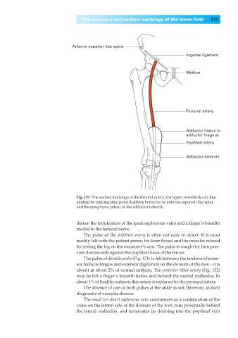
Anterior superior iliac spine
Inguinal ligament
Midline
Femoral artery
Adductor hiatus in
adductor magnus
Popliteal artery
Adductor tubercle
Fig. 153◊The surface markings of the femoral artery; the upper two-thirds of a line
joining the mid-inguinal point (halfway between the anterior superior iliac spine
and the symphysis pubis), to the adductor tubercle.
(hence the termination of the great saphenous vein) and a finger’s breadth
medial to the femoral nerve.
The pulse of the popliteal artery is often not easy to detect. It is most
readily felt with the patient prone, his knee flexed and his muscles relaxed
by resting the leg on the examiner’s arm. The pulse is sought by firm pres-
sure downwards against the popliteal fossa of the femur.
The pulse of dorsalis pedis (Fig. 151) is felt between the tendons of exten-
sor hallucis longus and extensor digitorum on the dorsum of the foot—it is
absent in about 2% of normal subjects. The posterior tibial artery (Fig. 152)
may be felt a finger’s breadth below and behind the medial malleolus. In
about 1% of healthy subjects this artery is replaced by the peroneal artery.
The absence of one or both pulses at the ankle is not, therefore, in itself
diagnostic of vascular disease.
The small (or short) saphenous vein commences as a continuation of the
veins on the lateral side of the dorsum of the foot, runs proximally behind
the lateral malleolus, and terminates by draining into the popliteal vein