
Page 248 - Clinical Anatomy
P. 248
ECA4 7/18/06 6:47 PM Page 233
The bones and joints of the lower limb 233
externally, you will note how the lateral and medial cartilages are respec-
tively sucked into the knee joint. If the flexed knee is forcibly abducted and
externally rotated, the medial cartilage will be drawn between, and then
split by, the grinding surfaces of the medial condyles of the femur and tibia.
This occurs when a footballer twists his flexed knee while running or when
a miner topples over in the crouched position while hewing coal in a
narrow seam. Asevere adduction and internal rotation strain may similarly
tear the lateral cartilage, but this injury is less common.
The knee ‘locks’ in this type of injury because the torn and displaced
segment of cartilage lodges between the condyles and prevents full exten-
sion of the knee.
The tibiofibular joints
The tibia and fibula are connected by:
1◊◊the superior tibiofibular joint, a synovial joint between the head of the
fibula and the lateral condyle of the tibia;
2◊◊the interosseous membrane, which is crossed by the anterior tibial vessels
above and pierced by the perforating branch of the peroneal artery below;
3◊◊the inferior tibiofibular joint, a fibrous joint, the only one in the limbs,
between the triangular areas of each bone immediately above the ankle
joint.
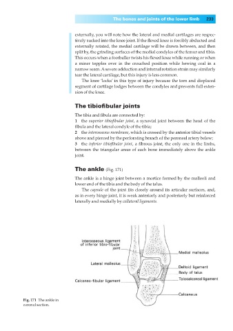
The ankle (Fig. 171)
The ankle is a hinge joint between a mortice formed by the malleoli and
lower end of the tibia and the body of the talus.
The capsule of the joint fits closely around its articular surfaces, and,
as in every hinge joint, it is weak anteriorly and posteriorly but reinforced
laterally and medially by collateral ligaments.
Fig. 171◊The ankle in
coronal section.