
Page 251 - Clinical Anatomy
P. 251
ECA4 7/18/06 6:47 PM Page 236
236 The lower limb
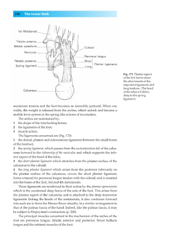
Fig. 173◊Plantar aspect
of the left foot to show
the attachments of the
important ligaments and
long tendons. (The head
of the talus is hidden,
deep to the spring
ligament).
maximum tension and the foot becomes an immobile pedestal. When one
walks, the weight is released from the arches, which unlock and become a
mobile lever system in the spring-like actions of locomotion.
The arches are maintained by:
1◊◊the shape of the interlocking bones;
2◊◊the ligaments of the foot;
3◊◊muscle action.
The ligaments concerned are (Fig. 173):
1◊◊the dorsal, plantar and interosseous ligaments between the small bones
of the forefoot;
2◊◊the spring ligament, which passes from the sustentaculum tali of the calca-
neus forward to the tuberosity of the navicular and which supports the infe-
rior aspect of the head of the talus;
3◊◊the short plantar ligament which stretches from the plantar surface of the
calcaneus to the cuboid;
4◊◊the long plantar ligament which arises from the posterior tuberosity on
the plantar surface of the calcaneus, covers the short plantar ligament,
forms a tunnel for peroneus longus tendon with the cuboid, and is inserted
into the bases of the 2nd, 3rd and 4th metatarsals.
These ligaments are reinforced in their action by the plantar aponeurosis
which is the condensed deep fascia of the sole of the foot. This arises from
the plantar aspect of the calcaneus and is attached to the deep transverse
ligaments linking the heads of the metatarsals; it also continues forward
into each toe to form the fibrous flexor sheaths, in a similar arrangement to
that of the palmar fascia of the hand. Indeed, like the palmar fascia, it may
be subject to Dupuytren’s contracture (p. 200).
The principal muscles concerned in the mechanism of the arches of the
foot are peroneus longus, tibialis anterior and posterior, flexor hallucis
longus and the intrinsic muscles of the foot.