
Page 250 - Clinical Anatomy
P. 250
ECA4 7/18/06 6:47 PM Page 235
The bones and joints of the lower limb 235
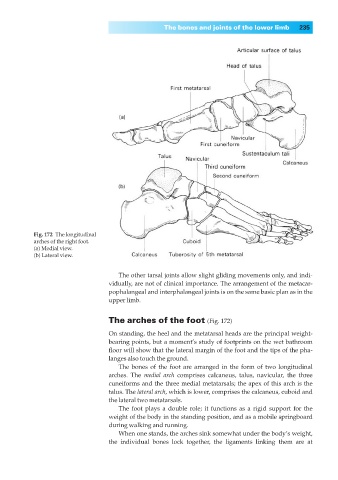
Fig. 172◊The longitudinal
arches of the right foot.
(a) Medial view.
(b) Lateral view.
The other tarsal joints allow slight gliding movements only, and indi-
vidually, are not of clinical importance. The arrangement of the metacar-
pophalangeal and interphalangeal joints is on the same basic plan as in the
upper limb.
The arches of the foot (Fig. 172)
On standing, the heel and the metatarsal heads are the principal weight-
bearing points, but a moment’s study of footprints on the wet bathroom
floor will show that the lateral margin of the foot and the tips of the pha-
langes also touch the ground.
The bones of the foot are arranged in the form of two longitudinal
arches. The medial arch comprises calcaneus, talus, navicular, the three
cuneiforms and the three medial metatarsals; the apex of this arch is the
talus. The lateral arch, which is lower, comprises the calcaneus, cuboid and
the lateral two metatarsals.
The foot plays a double role; it functions as a rigid support for the
weight of the body in the standing position, and as a mobile springboard
during walking and running.
When one stands, the arches sink somewhat under the body’s weight,
the individual bones lock together, the ligaments linking them are at