
Page 253 - Clinical Anatomy
P. 253
ECA4 7/18/06 6:47 PM Page 238
238 The lower limb
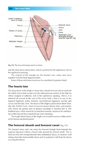
Fig. 174◊The femoral triangle and its contents.
and the deep fascia (fascia lata), which is pierced by the saphenous vein at
the saphenous opening.
The contents of the triangle are the femoral vein, artery and nerve
together with the deep inguinal nodes.
Some of these structures must now be considered in greater detail.
The fascia lata
The deep fascia of the thigh, or fascia lata, extends downwards to ensheath
the whole lower limb except over the subcutaneous surface of the tibia (to
whose margins it adheres), and at the saphenous opening. Above, it is
attached all around to the root of the lower limb — that is to say, to the
inguinal ligament, pubis, ischium, sacrotuberous ligament, sacrum and
coccyx and the iliac crest. The fascia of the thigh is particularly dense later-
ally (the iliotibial tract), where it receives tensor fasciae latae, and posteri-
orly, where the greater part of gluteus maximus is inserted into it. The
iliotibial tract, when tensed by its attached muscles, assists in the stabiliza-
tion of the hip and the extended knee when standing.
The tough lateral fascia of the thigh is an excellent source of this mater-
ial for hernia and dural repairs.
The femoral sheath and femoral canal (Fig. 175)
The femoral artery and vein enter the femoral triangle from beneath the
inguinal ligament within a fascial tube termed the femoral sheath. This is
derived from the extraperitoneal intra-abdominal fascia, its anterior wall
arising from the transversalis fascia and its posterior wall from the fascia
covering the iliacus.