
Page 254 - Clinical Anatomy
P. 254
ECA4 7/18/06 6:47 PM Page 239
Three important zones 239
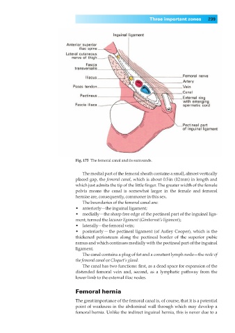
Fig. 175◊The femoral canal and its surrounds.
The medial part of the femoral sheath contains a small, almost vertically
placed gap, the femoral canal, which is about 0.5in (12mm) in length and
which just admits the tip of the little finger. The greater width of the female
pelvis means the canal is somewhat larger in the female and femoral
herniae are, consequently, commoner in this sex.
The boundaries of the femoral canal are:
•◊◊anteriorly—the inguinal ligament;
•◊◊medially—the sharp free edge of the pectineal part of the inguinal liga-
ment, termed the lacunar ligament (Gimbernat’s ligament);
•◊◊laterally—the femoral vein;
•◊◊posteriorly — the pectineal ligament (of Astley Cooper), which is the
thickened periosteum along the pectineal border of the superior pubic
ramus and which continues medially with the pectineal part of the inguinal
ligament.
The canal contains a plug of fat and a constant lymph node—the node of
the femoral canal or Cloquet’s gland.
The canal has two functions: first, as a dead space for expansion of the
distended femoral vein and, second, as a lymphatic pathway from the
lower limb to the external iliac nodes.
Femoral hernia
The great importance of the femoral canal is, of course, that it is a potential
point of weakness in the abdominal wall through which may develop a
femoral hernia. Unlike the indirect inguinal hernia, this is never due to a