
Page 255 - Clinical Anatomy
P. 255
ECA4 7/18/06 6:47 PM Page 240
240 The lower limb
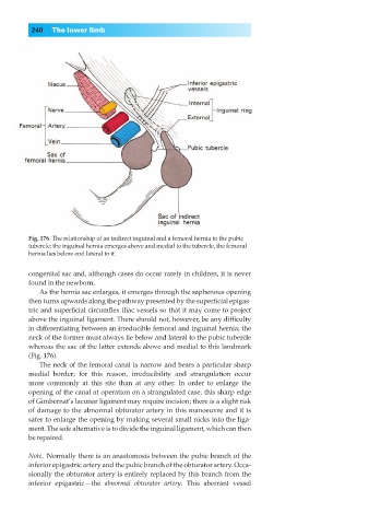
Fig. 176◊The relationship of an indirect inguinal and a femoral hernia to the pubic
tubercle; the inguinal hernia emerges above and medial to the tubercle, the femoral
hernia lies below and lateral to it.
congenital sac and, although cases do occur rarely in children, it is never
found in the newborn.
As the hernia sac enlarges, it emerges through the saphenous opening
then turns upwards along the pathway presented by the superficial epigas-
tric and superficial circumflex iliac vessels so that it may come to project
above the inguinal ligament. There should not, however, be any difficulty
in differentiating between an irreducible femoral and inguinal hernia; the
neck of the former must always lie below and lateral to the pubic tubercle
whereas the sac of the latter extends above and medial to this landmark
(Fig. 176).
The neck of the femoral canal is narrow and bears a particular sharp
medial border; for this reason, irreducibility and strangulation occur
more commonly at this site than at any other. In order to enlarge the
opening of the canal at operation on a strangulated case, this sharp edge
of Gimbernat’s lacunar ligament may require incision; there is a slight risk
of damage to the abnormal obturator artery in this manoeuvre and it is
safer to enlarge the opening by making several small nicks into the liga-
ment. The safe alternative is to divide the inguinal ligament, which can then
be repaired.
Note.◊Normally there is an anastomosis between the pubic branch of the
inferior epigastric artery and the pubic branch of the obturator artery. Occa-
sionally the obturator artery is entirely replaced by this branch from the
inferior epigastric —the abnormal obturator artery. This aberrant vessel