
Page 32 - Clinical Anatomy
P. 32
ECA1 7/18/06 6:31 PM Page 17
The thoracic cage 17
definitive position at the anterior part of the diaphragm. During this migra-
tion, the cervical myotomes and nerves contribute muscle and nerve
supply respectively, thus accounting for the long course of the phrenic
nerve (C3, 4 and 5) from the neck to the diaphragm.
With such a complex embryological story, one may be surprised to
know that congenital abnormalities of the diaphragm are unusual.
However, a number of defects may occur, giving rise to a variety of con-
genital herniae through the diaphragm. These may be:
1◊◊through the foramen of Morgagni; anteriorly between the xiphoid and
costal origins;
2◊◊through the foramen of Bochdalek— the pleuroperitoneal canal— lying
posteriorly;
3◊◊through a deficiency of the whole central tendon (occasionally such a
hernia may be traumatic in origin);
4◊◊through a congenitally large oesophageal hiatus.
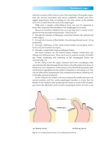
Far more common are the acquired hiatus herniae (subdivided into
sliding and rolling herniae). These are found in patients usually of middle
age where weakening and widening of the oesophageal hiatus has
occurred (Fig. 13).
In the sliding hernia the upper stomach and lower oesophagus slide
upwards into the chest through the lax hiatus when the patient lies down or
bends over; the competence of the cardia is often disturbed and peptic juice
can therefore regurgitate into the gullet in lying down or bending over. This
may be followed by oesophagitis with consequent heartburn, bleeding and,
eventually, stricture formation.
In the rolling hernia (which is far less common) the cardia remains in its
normal position and the cardio-oesophageal junction is intact, but the
fundus of the stomach rolls up through the hiatus in front of the oesopha-
gus, hence the alternative term of para-oesophageal hernia. In such a case
Fig. 13◊(a) Asliding hiatus hernia. (b) Arolling hiatus hernia.