
Page 296 - Clinical Anatomy
P. 296
ECA5 7/18/06 6:50 PM Page 281
The pharynx 281
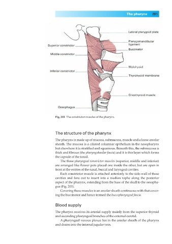
Fig. 203◊The constrictor muscles of the pharynx.
The structure of the pharynx
The pharynx is made up of mucosa, submucosa, muscle and a loose areolar
sheath. The mucosa is a ciliated columnar epithelium in the nasopharynx
but elsewhere it is stratified and squamous. Beneath this, the submucosa is
thick and fibrous (the pharyngobasilar fascia) and it is this layer which forms
the capsule of the tonsil.
The three pharyngeal constrictor muscles (superior, middle and inferior)
are arranged like flower pots placed one inside the other, but are open in
front at the entries of the nasal, buccal and laryngeal cavities.
Each constrictor muscle is attached anteriorly to the side-wall of these
cavities and fans out to insert into a median raphe along the posterior
aspect of the pharynx, extending from the base of the skull to the oesopha-
gus (Fig. 203).
Covering these muscles is an areolar sheath continuous with that cover-
ing the buccinator and hence termed the buccopharyngeal fascia.
Blood supply
The pharynx receives its arterial supply mainly from the superior thyroid
and ascending pharyngeal branches of the external carotid.
A pharyngeal venous plexus lies in the areolar sheath of the pharynx
and drains into the internal jugular vein.