
Page 292 - Clinical Anatomy
P. 292
ECA5 7/18/06 6:50 PM Page 277
The pharynx 277
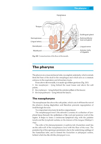
Hyoglossus
Tongue
Sublingual gland
Genioglossus Submandibular
duct and gland
Lingual artery
Geniohyoid Lingual nerve
Mylohyoid Anterior belly
of digastric
Fig. 200◊Coronal section of the floor of the mouth.
The pharynx
The pharynx is a musculofascial tube, incomplete anteriorly, which extends
from the base of the skull to the oesophagus and which acts as a common
entrance to the respiratory and alimentary tracts.
From above downwards, it is made up of three portions (Fig. 201):
1◊◊the nasopharynx — lying behind the nasal fossae and above the soft
palate;
2◊◊the oropharynx—lying behind the anterior pillars of the fauces;
3◊◊the laryngopharynx—lying behind the larynx.
The nasopharynx
The nasopharynx lies above the soft palate, which cuts it off from the rest of
the pharynx during deglutition and therefore prevents regurgitation of
food through the nose.
Two important structures lie in this compartment.
The nasopharyngeal tonsil (‘the adenoids’) consists of a collection of lym-
phoid tissue beneath the epithelium of the roof and posterior wall of this
region. It helps to form a continuous lymphoid ring with the palatine
tonsils and the lymphoid nodules on the dorsum of the tongue (Waldeyer’s
ring).
The orifice of the pharyngotympanic or auditory tube (Eustachian canal) lies
on the side-wall of the nasopharynx level with the floor of the nose. The
posterior lip of this opening is prominent, due to the underlying cartilage of
the Eustachian tube, and is termed the Eustachian or pharyngeal cushion,
behind which lies the slit-like pharyngeal recess.