
Page 301 - Clinical Anatomy
P. 301
ECA5 7/18/06 6:50 PM Page 286
286 The head and neck
Epiglottis
Hyo-epiglottic
ligament
Lateral thyrohyoid
ligament Hyoid
Median thyrohyoid
ligament
Arytenoid cartilage
Vestibular fold
Vocal and Sinus of larynx
muscular processes
of arytenoid Vocal fold
Cricovocal membrane
Cricothyroid ligament
Facet on cricoid
for inferior horn
of thyroid cartilage
Cricotracheal ligament
(a)
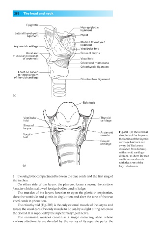
Fig. 206◊(a) The internal
structure of the larynx—
the lamina of the thyroid
cartilage has been cut
away. (b) The larynx
dissected from behind,
with cricoid cartilage
divided, to show the true
and false vocal cords
with the sinus of the
(b) larynx between.
3◊◊the subglottic compartment between the true cords and the first ring of
the trachea.
On either side of the larynx the pharynx forms a recess, the piriform
fossa, in which swallowed foreign bodies tend to lodge.
The muscles of the larynx function to open the glottis in inspiration,
close the vestibule and glottis in deglutition and alter the tone of the true
vocal cords in phonation.
The cricothyroid (Fig. 203) is the only external muscle of the larynx and
tenses the vocal cord (the only muscle to do so), by a slight tilting action on
the cricoid. It is supplied by the superior laryngeal nerve.
The remaining muscles constitute a single encircling sheet whose
various attachments are denoted by the names of its separate parts: the