
Page 293 - Clinical Anatomy
P. 293
ECA5 7/18/06 6:50 PM Page 278
278 The head and neck
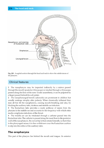
Fig. 201◊Asagittal section through the head and neck to show the subdivisions of
the pharynx.
Clinical features
1◊◊The nasopharynx may be inspected indirectly by a mirror passed
through the mouth (posterior rhinoscopy) or studied through a rhinoscope
passed along the floor of the nose. Under anaesthesia, it can be palpated by
a finger passed behind the soft palate.
2◊◊The nasopharyngeal tonsils (adenoids) are prominent in children but
usually undergo atrophy after puberty. When chronically inflamed they
may all but fill the nasopharynx, causing mouth-breathing and also, by
blocking the auditory tube, deafness and middle ear infection.
3◊◊The Eustachian tube provides a ready pathway of sepsis from the
pharynx to the middle ear and accounts for the frequency with which otitis
media complicates infections of the throat.
4◊◊The middle ear can be intubated through a catheter passed into the
Eustachian tube. The catheter is passed along the nasal floor to the posterior
wall of the nasopharynx. Its curved tip is then rotated laterally so that it lies
in the pharyngeal recess; it is then withdrawn over the Eustachian cushion
to slip into the orifice of the auditory tube.
The oropharynx
This part of the pharynx lies behind the mouth and tongue. Its anterior