
Page 294 - Clinical Anatomy
P. 294
ECA5 7/18/06 6:50 PM Page 279
The pharynx 279
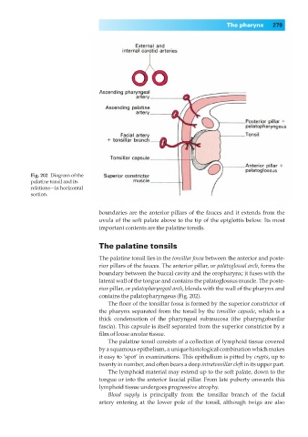
Fig. 202◊Diagram of the
palatine tonsil and its
relations—in horizontal
section.
boundaries are the anterior pillars of the fauces and it extends from the
uvula of the soft palate above to the tip of the epiglottis below. Its most
important contents are the palatine tonsils.
The palatine tonsils
The palatine tonsil lies in the tonsillar fossa between the anterior and poste-
rior pillars of the fauces. The anterior pillar, or palatoglossal arch, forms the
boundary between the buccal cavity and the oropharynx; it fuses with the
lateral wall of the tongue and contains the palatoglossus muscle. The poste-
rior pillar, or palatopharyngeal arch, blends with the wall of the pharynx and
contains the palatopharyngeus (Fig. 202).
The floor of the tonsillar fossa is formed by the superior constrictor of
the pharynx separated from the tonsil by the tonsillar capsule, which is a
thick condensation of the pharyngeal submucosa (the pharyngobasilar
fascia). This capsule is itself separated from the superior constrictor by a
film of loose areolar tissue.
The palatine tonsil consists of a collection of lymphoid tissue covered
by a squamous epithelium, a unique histological combination which makes
it easy to ‘spot’ in examinations. This epithelium is pitted by crypts, up to
twenty in number, and often bears a deep intratonsillar cleft in its upper part.
The lymphoid material may extend up to the soft palate, down to the
tongue or into the anterior faucial pillar. From late puberty onwards this
lymphoid tissue undergoes progressive atrophy.
Blood supply is principally from the tonsillar branch of the facial
artery entering at the lower pole of the tonsil, although twigs are also