
Page 302 - Clinical Anatomy
P. 302
ECA5 7/18/06 6:50 PM Page 287
The larynx 287
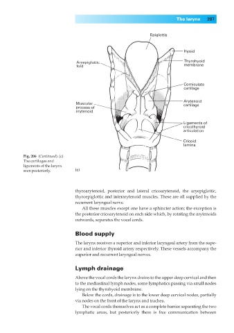
Fig. 206◊(Continued). (c)
The cartilages and
ligaments of the larynx
seen posteriorly. (c)
thyroarytenoid, posterior and lateral cricoarytenoid, the aryepiglottic,
thyroepiglottic and interarytenoid muscles. These are all supplied by the
recurrent laryngeal nerve.
All these muscles except one have a sphincter action; the exception is
the posterior cricoarytenoid on each side which, by rotating the arytenoids
outwards, separates the vocal cords.
Blood supply
The larynx receives a superior and inferior laryngeal artery from the supe-
rior and inferior thyroid artery respectively. These vessels accompany the
superior and recurrent laryngeal nerves.
Lymph drainage
Above the vocal cords the larynx drains to the upper deep cervical and then
to the mediastinal lymph nodes, some lymphatics passing via small nodes
lying on the thyrohyoid membrane.
Below the cords, drainage is to the lower deep cervical nodes, partially
via nodes on the front of the larynx and trachea.
The vocal cords themselves act as a complete barrier separating the two
lymphatic areas, but posteriorly there is free communication between