
Page 305 - Clinical Anatomy
P. 305
ECA5 7/18/06 6:50 PM Page 290
290 The head and neck
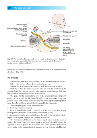
Fig. 208◊The parotid and its surrounds in a schematic horizontal section—the facial
nerve is the most superficial of the structures traversing the gland. (The line of
section is shown in the inset head.)
mandible and sternocleidomastoid and overflowing both these bounding
structures (Fig. 208).
Relations
•◊◊Above—lie the external auditory meatus and temporomandibular joint.
•◊◊Below—it overflows the posterior belly of digastric.
•◊◊Anteriorly—it overflows the mandible with the overlying masseter.
•◊◊Medially — lies the styloid process and its muscles separating the
parotid from the internal jugular vein, internal carotid artery, last four
cranial nerves and the lateral wall of the pharynx.
The gland itself is enclosed in a split in the investing fascia, lying both
on and below which are the parotid lymph nodes. Antero-inferiorly, this
parotid fascia is thickened and is the only structure separating the parotid
from the submandibular gland (the stylomandibular ligament).
Traversing the gland (from without in) are:
1◊◊the facial nerve (see below);
2◊◊the retromandibular (posterior facial) vein, formed by the junction of
the superficial temporal and maxillary veins (See Fig. 216);
3◊◊the external carotid artery, dividing at the neck of the mandible into its
superficial temporal and maxillary terminal branches.
The parotid duct (of Stensen) is 2in (5cm) long. It arises from the anterior
part of the gland, runs over the masseters a finger’s breadth below the
zygomatic arch to pierce the buccinator and open opposite the second
upper molar tooth. Inspect this in the mirror in your own mouth. The duct