Page 326 - Clinical Anatomy
P. 326

ECA5  7/18/06  6:51 PM  Page 311


                                        The surface anatomy and surface markings of the head   311


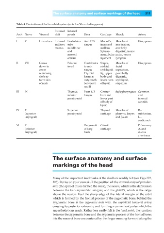
                  Table 4◊Derivatives of the branchial system (note the 5th arch disappears).

                                         External  Internal
                  Arch Nerve    Visceral  cleft  pouch    Floor     Cartilage  Muscle     Artery
                  I    V       Lower face External  Eustachian  Antr 2/3   Meckel’s,   Muscles of  Disappears
                                         auditory tube,   tongue    incus and  mastication,
                                         meatus  middle ear         malleus  antr belly
                                                and                 Spheno-  digastric, tensor
                                                mastoid             mandibular palati, tensor
                                                antrum              ligament  tympani
                  II   VII     Grows            Palatine  Contributes Stapes,  Muscles of  Disappears
                               down to          tonsil    to antr   styloid,  facial
                               cover                      tongue    stylohyoid expression,
                               remaining                  Thyroid   lig, upper  postr belly
                               clefts to                  forms as   body and   digastric,
                               form skin                  outgrowth   lesser horn  stylohyoid,
                               of neck                    between I   of hyoid  stapedius
                                                          and II
                  III  IX                       Thymus,   Postr 1/3   Greater    Stylopharyngeus Common
                                                inferior  tongue    horn and              and
                                                parathyroid         lower part            internal
                                                                    of body of            carotids
                                                                    hyoid
                  IV   X                        Superior            Thyroid  Muscles of   R—
                       (superior                parathyroid         cartilage  pharynx, larynx  subclavian,
                       laryngeal)                                            and palate   L—
                                                                                          aortic arch
                  VI   X                                  Outgrowth Cricoid               Pulmonary
                       (inferior                          of lung   cartilage             A. and
                       laryngeal)                         buds                            ductus
                                                                                          arteriosus


                                       The surface anatomy and surface

                                       markings of the head


                                       Many of the important landmarks of the skull are readily felt (see Figs 222,
                                       223). Revise on your own skull the position of: the external occipital protuber-
                                       ance (the apex of this is termed the inion), the nasion, which is the depression
                                        between the two supraorbital margins, and the glabella, which is the ridge
                                       above the nasion. Feel the sharp edge of the lateral margin of the orbit
                                       which is formed by the frontal process of the zygomatic bone; behind the
                                       zygomatic bone is the  zygomatic arch with the  superficial temporal artery
                                       crossing its posterior extremity and forming a convenient pulse which the
                                       anaesthetist can reach. Rather less easily felt is the jugal point, the junction
                                       between the zygomatic bone and the zygomatic process of the frontal bone;
                                       it is the mass of bone encountered by the finger running forward along the
   321   322   323   324   325   326   327   328   329   330   331