
Page 331 - Clinical Anatomy
P. 331
ECA5 7/18/06 6:51 PM Page 316
316 The head and neck
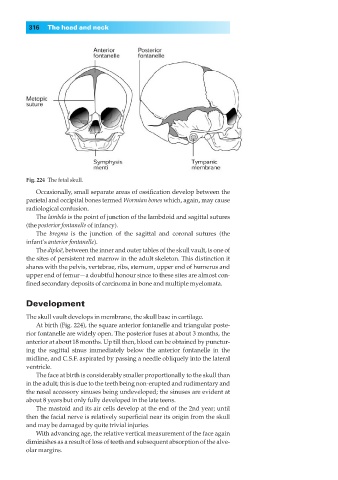
Fig. 224◊The fetal skull.
Occasionally, small separate areas of ossification develop between the
parietal and occipital bones termed Wormian bones which, again, may cause
radiological confusion.
The lambda is the point of junction of the lambdoid and sagittal sutures
(the posterior fontanelle of infancy).
The bregma is the junction of the sagittal and coronal sutures (the
infant’s anterior fontanelle).
The diploë, between the inner and outer tables of the skull vault, is one of
the sites of persistent red marrow in the adult skeleton. This distinction it
shares with the pelvis, vertebrae, ribs, sternum, upper end of humerus and
upper end of femur—a doubtful honour since to these sites are almost con-
fined secondary deposits of carcinoma in bone and multiple myelomata.
Development
The skull vault develops in membrane, the skull base in cartilage.
At birth (Fig. 224), the square anterior fontanelle and triangular poste-
rior fontanelle are widely open. The posterior fuses at about 3 months, the
anterior at about 18 months. Up till then, blood can be obtained by punctur-
ing the sagittal sinus immediately below the anterior fontanelle in the
midline, and C.S.F. aspirated by passing a needle obliquely into the lateral
ventricle.
The face at birth is considerably smaller proportionally to the skull than
in the adult; this is due to the teeth being non-erupted and rudimentary and
the nasal accessory sinuses being undeveloped; the sinuses are evident at
about 8 years but only fully developed in the late teens.
The mastoid and its air cells develop at the end of the 2nd year; until
then the facial nerve is relatively superficial near its origin from the skull
and may be damaged by quite trivial injuries.
With advancing age, the relative vertical measurement of the face again
diminishes as a result of loss of teeth and subsequent absorption of the alve-
olar margins.