
Page 328 - Clinical Anatomy
P. 328
ECA5 7/18/06 6:51 PM Page 313
The scalp 313
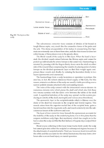
Fig. 221◊The layers of the
scalp.
The subcutaneous connective tissue consists of lobules of fat bound in
tough fibrous septa, very much like the connective tissue of the palm and
the sole. This dense encapsulation of fat makes it unsurprising that lipo-
mata are extremely rare at these three sites, and also that excess fat does not
collect in any of these places even in the grossly obese.
The blood vessels of the scalp lie in this layer. When the head is lacer-
ated, the divided vessels retract between the fibrous septa and cannot be
picked up individually by artery forceps in the usual way. Haemorrhage is
arrested by pressing with the fingers firmly down on to the skull on either
side of the wound (thus compressing the vessels), by placing series of artery
forceps on the divided aponeurotic layer so that their weight again com-
presses these vessels and, finally, by suturing the laceration firmly in two
layers (aponeurotic and cutaneous).
The haemorrhage from a scalp laceration or operation is profuse; this
area has, in fact, the richest cutaneous blood supply of the body. For this
reason, extensive avulsions of the scalp are usually viable providing even a
narrow pedicle remains attached to the surrounding tissues.
The veins of the scalp connect with the intracranial venous sinuses via
numerous emissary veins which pierce the skull and which also link these
two venous systems with the diploic veins between the tables of the skull
vault. A superficial infection of the scalp may spread via this system pro-
ducing an osteitis of the skull, meningitis and venous sinus thrombosis.
The aponeurotic layer is the occipitofrontalis which is fibrous over the
dome of the skull but muscular in the occipital and frontal regions. This
muscle arises from the superior nuchal line of the occipital bone, gains a
fascial insertion into the zygomatic arch, and inserts anteriorly into the sub-
cutaneous tissues of the eyebrows and nose.
The layer of loose connective tissue beneath the aponeurosis accounts for
the mobility of the scalp on the underlying bone; it is in this plane that the
surgeon mobilizes scalp flaps, that machinery which has caught on to the
hair avulses the scalp and that the Red Indians of bygone days scalped their
victims.
Blood or pus collecting in this loose tissue tracks freely under the scalp
but cannot pass into either the occipital or subtemporal regions because of
the attachments of occipitofrontalis. Fluid can, however, track forward into
the orbits and this accounts for the orbital haematoma that may form a few
hours after a severe head injury or cranial operation.