Page 36 - Clinical Anatomy
P. 36

ECA1  7/18/06  6:31 PM  Page 21


                                                                   The lower respiratory tract  21


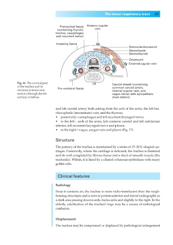
                                          Pretracheal fascia  Anterior jugular
                                        (containing thyroid,  vein
                                        trachea, oesophagus
                                        and recurrent nerve)

                                        Investing fascia
                                                                                 Sternocleidomastoid
                                                                                 Sternohyoid
                                                                                 Sternothyroid
                                                                                 Omohyoid
                                                                                 External jugular vein


                  Fig. 16◊The cervical part                 C6         Carotid sheath (containing
                  of the trachea and its  Pre-vertebral fascia         common carotid artery,
                  environs in transverse                               internal jugular vein, and
                  section (through the 6th                             vagus nerve) with sympathetic
                  cervical vertebra).                                  chain behind


                                       and left carotid artery, both arising from the arch of the aorta, the left bra-
                                       chiocephalic (innominate) vein, and the thymus;
                                       •◊◊posteriorly—oesophagus and left recurrent laryngeal nerve;
                                       •◊◊to the left — arch of the aorta, left common carotid and left subclavian
                                       arteries, left recurrent laryngeal nerve and pleura;
                                       •◊◊to the right—vagus, azygos vein and pleura (Fig. 17).

                                       Structure

                                       The patency of the trachea is maintained by a series of 15–20 U-shaped car-
                                       tilages. Posteriorly, where the cartilage is deficient, the trachea is flattened
                                       and its wall completed by fibrous tissue and a sheet of smooth muscle (the
                                       trachealis). Within, it is lined by a ciliated columnar epithelium with many
                                       goblet cells.


                                         Clinical features

                                       Radiology

                                       Since it contains air, the trachea is more radio-translucent than the neigh-
                                       bouring structures and is seen in posteroanterior and lateral radiographs as
                                       a dark area passing downwards, backwards and slightly to the right. In the
                                       elderly, calcification of the tracheal rings may be a source of radiological
                                       confusion.

                                       Displacement

                                       The trachea may be compressed or displaced by pathological enlargement
   31   32   33   34   35   36   37   38   39   40   41