
Page 41 - Clinical Anatomy
P. 41
ECA1 7/18/06 6:31 PM Page 26
26 The Thorax
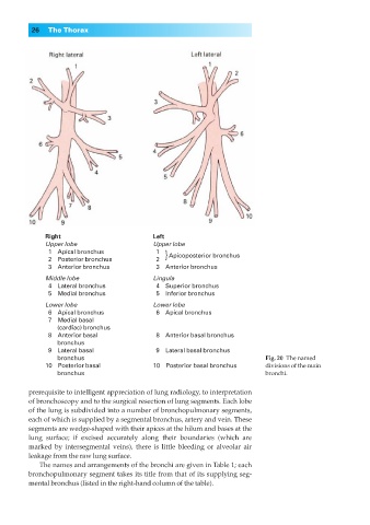
Right Left
Upper lobe Upper lobe
◊1◊◊Apical bronchus ◊1◊◊ } Apicoposterior bronchus
◊2◊◊Posterior bronchus ◊2◊◊
◊3◊◊Anterior bronchus ◊3◊◊Anterior bronchus
Middle lobe Lingula
◊4◊◊Lateral bronchus ◊4◊◊Superior bronchus
◊5◊◊Medial bronchus ◊5◊◊Inferior bronchus
Lower lobe Lower lobe
◊6◊◊Apical bronchus ◊6◊◊Apical bronchus
◊7◊◊Medial basal
◊◊◊◊(cardiac) bronchus
◊8◊◊Anterior basal ◊8◊◊Anterior basal bronchus
◊◊◊◊bronchus
◊9◊◊Lateral basal ◊9◊◊Lateral basal bronchus
◊◊◊◊bronchus Fig. 20◊The named
10◊◊Posterior basal 10◊◊Posterior basal bronchus divisions of the main
◊◊◊◊bronchus bronchi.
prerequisite to intelligent appreciation of lung radiology, to interpretation
of bronchoscopy and to the surgical resection of lung segments. Each lobe
of the lung is subdivided into a number of bronchopulmonary segments,
each of which is supplied by a segmental bronchus, artery and vein. These
segments are wedge-shaped with their apices at the hilum and bases at the
lung surface; if excised accurately along their boundaries (which are
marked by intersegmental veins), there is little bleeding or alveolar air
leakage from the raw lung surface.
The names and arrangements of the bronchi are given in Table 1; each
bronchopulmonary segment takes its title from that of its supplying seg-
mental bronchus (listed in the right-hand column of the table).