
Page 40 - Clinical Anatomy
P. 40
ECA1 7/18/06 6:31 PM Page 25
The lower respiratory tract 25
Blood supply
Mixed venous blood is returned to the lungs by the pulmonary arteries; the
air passages are themselves supplied by the bronchial arteries, which are
small branches of the descending aorta. The bronchial arteries, although
small, are of great clinical importance. They maintain the blood supply to
the lung parenchyma after pulmonary embolism, so that, if the patient
recovers, lung function returns to normal.
The superior and inferior pulmonary veins return oxygenated blood to the
left atrium, while the bronchial veins drain into the azygos system.
Lymphatic drainage
The lymphatics of the lung drain centripetally from the pleura towards the
hilum. From the bronchopulmonary lymph nodes in the hilum, efferent lymph
channels pass to the tracheobronchial nodes at the bifurcation of the trachea,
thence to the paratracheal nodes and the mediastinal lymph trunks to drain
usually directly into the brachiocephalic veins or, rarely, indirectly via the
thoracic or right lymphatic duct.
Nerve supply
The pulmonary plexuses derive fibres from both the vagi and the sympa-
thetic trunk. They supply efferents to the bronchial musculature (sympa-
thetic bronchodilator fibres) and receive afferents from the mucous
membrane of the bronchioles and from the alveoli.
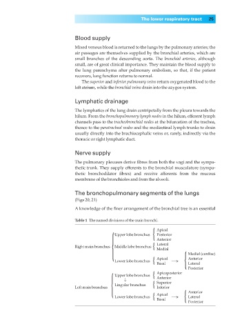
The bronchopulmonary segments of the lungs
(Figs 20, 21)
A knowledge of the finer arrangement of the bronchial tree is an essential
Table 1◊The named divisions of the main bronchi. Apical
{ Upper lobe bronchus { Posterior
Anterior
Middle lobe bronchus {
Lateral
Right main bronchus Medial
Lower lobe bronchus { Apical → { Medial (cardiac)
Anterior
Basal Lateral
Posterior
Upper lobe bronchus {
Apicoposterior
{ Lingular bronchus { Anterior
↓
Superior
Left main bronchus Inferior Anterior
Lower lobe bronchus { Apical → { Lateral
Basal
Posterior