
Page 378 - Clinical Anatomy
P. 378
ECA6 7/18/06 6:54 PM Page 363
The brain 363
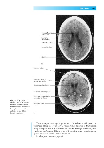
Fig. 252◊(a) CT scan of
skull through the level of
the bodies of the lateral
ventricles. (b) CT scan cut
through the level of the
anterior horns of the
lateral ventricles.
6◊◊The meningeal coverings, together with the subarachnoid space, are
prolonged along the optic nerve. Raised C.S.F. pressure is transmitted
along this space and may compress the venous drainage of the eye, thus
producing papilloedema. This swelling of the optic disc can be detected by
ophthalmoscopic examination of the fundus.
7◊◊Lumbar puncture—see page 338.