
Page 381 - Clinical Anatomy
P. 381
ECA6 7/18/06 6:54 PM Page 366
366 The central nervous system
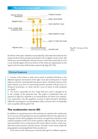
Fig. 255◊The layers of the
retina.
the fibres of the optic radiation sweep laterally, and some forwards into the
temporal lobe before passing backwards to the occipital visual cortex (the
striate area surrounding the calcarine fissure) where they terminate in such
a way that the upper and lower halves of the retina are represented on the
upper and lower lips of the fissure respectively (Figs 247, 256).
Clinical features
1◊◊Lesions of the retina or optic nerve result in ipsilateral blindness in the
affected segment, but lesions of the optic tract and central parts of visual
pathway result in contralateral homonymous defects. Similarly, lesions of the
optic chiasma (e.g. from an expanding pituitary tumour) will give rise to a
bitemporal hemianopia, i.e. there will be a loss of vision in both temporal
eye-fields.
2◊◊The lesion responsible for the Argyll Robertson pupil is thought to be
in the vicinity of the pretectal area. The pupil is constricted, does not
respond to light but responds to accommodation, but there is no satisfac-
tory explanation why the pupillary reaction to light should be abolished
while the convergence–accommodation reflex is preserved. It is classically
seen in syphilis affecting the CNS.
The oculomotor nerve (III)
In addition to supplying most of the extrinsic eye muscles, the oculomotor
nerve conveys the preganglionic parasympathetic fibres for the sphincter of
the pupil via the ciliary ganglion. Its nucleus of origin lies in the floor of the
cerebral aqueduct at the level of the superior colliculus (Fig. 245) and con-
sists essentially of two components: the somatic efferent nucleus, which sup-
plies the ocular muscles, and the Edinger–Westphal nucleus from which the
preganglionic parasympathetic fibres are derived.
From these nuclei, fibres pass vertically through the midbrain tegmen-