
Page 383 - Clinical Anatomy
P. 383
ECA6 7/18/06 6:54 PM Page 368
368 The central nervous system
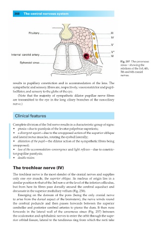
Fig. 257◊The cavernous
sinus—showing the
relations of the 3rd, 4th,
5th and 6th cranial
nerves.
results in pupillary constriction and in accommodation of the lens. The
sympathetic and sensory fibres are, respectively, vasoconstrictor and pupil-
lodilator, and sensory to the globe of the eye.
(Note that the majority of sympathetic dilator pupillae nerve fibres
are transmitted to the eye in the long ciliary branches of the nasociliary
nerve.)
Clinical features
Complete division of the 3rd nerve results in a characteristic group of signs:
•◊◊ptosis—due to paralysis of the levator palpebrae superioris;
•◊◊a divergent squint—due to the unopposed action of the superior oblique
and lateral rectus muscles, rotating the eyeball laterally;
•◊◊dilatation of the pupil—the dilator action of the sympathetic fibres being
unopposed;
•◊◊loss of the accommodation–convergence and light reflexes— due to constric-
tor pupillae paralysis;
•◊◊double vision.
The trochlear nerve (IV)
The trochlear nerve is the most slender of the cranial nerves and supplies
only one eye muscle, the superior oblique. Its nucleus of origin lies in a
similar position to that of the 3rd nerve at the level of the inferior colliculus,
but from here its fibres pass dorsally around the cerebral aqueduct and
decussate in the superior medullary vellum (Fig. 258).
Emerging on the dorsum of the pons (being the only cranial nerve
to arise from the dorsal aspect of the brainstem), the nerve winds round
the cerebral peduncle and then passes forwards between the superior
cerebellar and posterior cerebral arteries to pierce the dura. It then runs
forwards in the lateral wall of the cavernous sinus (Fig. 257) between
the oculomotor and ophthalmic nerves to enter the orbit through the supe-
rior orbital fissure, lateral to the tendinous ring from which the recti take