
Page 384 - Clinical Anatomy
P. 384
ECA6 7/18/06 6:54 PM Page 369
The cranial nerves 369
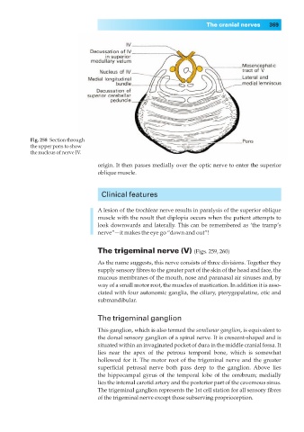
Fig. 258◊Section through
the upper pons to show
the nucleus of nerve IV.
origin. It then passes medially over the optic nerve to enter the superior
oblique muscle.
Clinical features
A lesion of the trochlear nerve results in paralysis of the superior oblique
muscle with the result that diplopia occurs when the patient attempts to
look downwards and laterally. This can be remembered as ‘the tramp’s
nerve”—it makes the eye go “down and out”!
The trigeminal nerve (V) (Figs. 259, 260)
As the name suggests, this nerve consists of three divisions. Together they
supply sensory fibres to the greater part of the skin of the head and face, the
mucous membranes of the mouth, nose and paranasal air sinuses and, by
way of a small motor root, the muscles of mastication. In addition it is asso-
ciated with four autonomic ganglia, the ciliary, pterygopalatine, otic and
submandibular.
The trigeminal ganglion
This ganglion, which is also termed the semilunar ganglion, is equivalent to
the dorsal sensory ganglion of a spinal nerve. It is crescent-shaped and is
situated within an invaginated pocket of dura in the middle cranial fossa. It
lies near the apex of the petrous temporal bone, which is somewhat
hollowed for it. The motor root of the trigeminal nerve and the greater
superficial petrosal nerve both pass deep to the ganglion. Above lies
the hippocampal gyrus of the temporal lobe of the cerebrum; medially
lies the internal carotid artery and the posterior part of the cavernous sinus.
The trigeminal ganglion represents the 1st cell station for all sensory fibres
of the trigeminal nerve except those subserving proprioception.