
Page 44 - Clinical Anatomy
P. 44
ECA1 7/18/06 6:31 PM Page 29
The mediastinum 29
great vessels and the base with the central tendon of the diaphragm. Anteri-
orly it is related to the body of the sternum, to which it is attached by the
sternopericardial ligament. The 3rd–6th costal cartilages and the anterior
borders of the lungs; posteriorly, to the oesophagus, descending aorta, and
vertebra T5–T8, and on either side to the roots of the lungs, the mediastinal
pleura and the phrenic nerves.
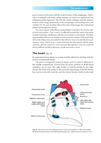
The inner aspect of the fibrous pericardium is lined by the parietal layer
of serous pericardium. This, in turn, is reflected around the roots of the great
vessels to become continuous with the visceral layer or epicardium. The lines
of pericardial reflexion are marked on the posterior surface of the heart (Fig.
23) by the oblique sinus, bounded by the inferior vena cava and the four pul-
monary veins, which form a recess between the left atrium and the peri-
cardium, and the transverse sinus between the superior vena cava and left
atrium behind and the pulmonary trunk and aorta in front.
The heart (Fig. 24)
Its great importance means no excuse need be offered for dealing with the
heart in considerable detail.
The heart is irregularly conical in shape, and it is placed obliquely in
the middle mediastinum. Viewed from the front, portions of all the heart
chambers can be seen. The right border is formed entirely by the right
atrium, the left border partly by the auricular appendage of the left atrium
but mainly by the left ventricle, and the inferior border chiefly by the right
Fig. 23◊The transverse and oblique sinuses of the pericardium. In this illustration
the heart has been removed from the pericardial sac, which is seen in anterior view.