
Page 49 - Clinical Anatomy
P. 49
ECA1 7/18/06 6:31 PM Page 34
34 The Thorax
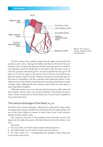
Fig. 27◊The coronary
arteries. (Dotted vessels
lie posteriorly.)
The left coronary artery, which is larger than the right, rises from the left
posterior aortic sinus. Passing first behind and then to the left of the pul-
monary trunk, it reaches the left part of atrioventricular groove in which it
runs laterally round the left border of the heart as the circumflex artery to
reach the posterior interatrial groove. Its most important branch, given off
about 2 cm from its origin, is the anterior interventricular artery which sup-
plies the anterior aspect of both ventricles and passes around the apex of
the heart to anastomose with the posterior interventricular branch of the
right coronary. Note that the sinuatrial node is usually supplied by the right
coronary artery, although the left coronary artery takes over this duty in
about one-third of subjects.
Although anastomoses occur between the terminations of the right and
left coronary arteries, these are usually inefficient. Thrombosis in one or
other of these vessels leads to death of the area of heart muscle supplied (a
myocardial infarction).
The venous drainage of the heart (Fig. 28)
The bulk of the venous drainage of the heart is achieved by veins which
accompany the coronary arteries and which open into the right atrium. The
rest of the blood drains by means of small veins (venae cordis minimae)
directly into the cardiac cavity.
The coronary sinus lies in the posterior atrioventricular groove and
opens into the right atrium just to the left of the mouth of the inferior vena
cava.
It receives:
1◊◊the great cardiac vein in the anterior interventricular groove;
2◊◊the middle cardiac vein the inferior interventricular groove;
3◊◊the small cardiac vein — accompanying the marginal artery along the
lower border of the heart;