
Page 46 - Clinical Anatomy
P. 46
ECA1 7/18/06 6:31 PM Page 31
The mediastinum 31
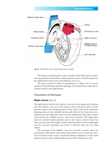
Fig. 25◊The interior of the right atrium and ventricle.
The inferior or diaphragmatic surface consists of the right and left ventri-
cles separated by the posterior interventricular groove and the portion of
the right atrium which receives the inferior vena cava.
The base or posterior surface is quadrilateral in shape and is formed
mainly by the left atrium with the openings of the pulmonary veins and, to
a lesser extent, by the right atrium.
Chambers of the heart
Right atrium (Fig. 25)
The right atrium receives the superior vena cava in its upper and posterior
part, the inferior vena cava and coronary sinus in its lower part, and the
anterior cardiac vein (draining much of the front of the heart) anteriorly.
Running more or less vertically downwards between the venae cavae is a
distinct muscular ridge, the crista terminalis (indicated on the outer surface
of the atrium by a shallow groove— the sulcus terminalis). This ridge sepa-
rates the smooth-walled posterior part of the atrium, derived from the
sinus venosus, from the rough-walled anterior portion which is prolonged
into the auricular appendage and which is derived from the true fetal
atrium.
The openings of the inferior vena cava and the coronary sinus are
guarded by rudimentary valves; that of the inferior vena cava being contin-
uous with the annulus ovalis around the shallow depression on the atrial
septum, the fossa ovalis, which marks the site of the fetal foramen ovale.