
Page 50 - Clinical Anatomy
P. 50
ECA1 7/18/06 6:31 PM Page 35
The mediastinum 35
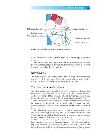
Fig. 28◊The coronary veins. (Dotted vessels lie posteriorly.)
4◊◊the oblique vein — descends obliquely on the posterior aspect of the left
atrium.
The anterior cardiac veins (up to three or four in number) cross the ante-
rior atrioventricular groove, drain much of the anterior surface of the heart
and open directly into the right atrium.
Nerve supply
The nerve supply of the heart is derived from the vagus (cardio-inhibitor)
and the cervical and upper 5 thoracic sympathetic ganglia (cardio-
accelerator) by way of superficial and deep cardiac plexuses.
The development of the heart
The primitive heart is a single tube which soon shows grooves demarcating
the sinus venosus, atrium, ventricle and bulbus cordis from behind forwards.
As this tube enlarges it kinks so that its caudal end, receiving venous blood,
comes to lie behind its cephalic end with its emerging arteries (Fig. 29).
The sinus venosus later absorbs into the atrium and the bulbus becomes
incorporated into the ventricle so that, in the fully developed heart, the
atria and great veins come to lie posterior to the ventricles and the roots of
the great arteries.
The boundary tissue between the primitive single atrial cavity
and single ventricle grows out as a dorsal and a ventral endocardial cushion
which meet in the midline, thus dividing the common atrio-ventricular
orifice into a right (tricuspid) and left (mitral) orifice.
The division of the primitive atrium into two is a complicated process
but an important one in the understanding of congenital septal defects
(Fig. 30). Apartition, the septum primum, grows downwards from the poste-
rior and superior walls of the primitive common atrium to fuse with the