
Page 78 - Clinical Anatomy
P. 78
ECA2 7/18/06 6:42 PM Page 63
The fasciae and muscles of the abdominal wall 63
This is used, for example, on the left in removing growths of the upper
stomach or lower oesophagus and on the right in resection of the right lobe
of the liver.
Paracentesis abdominis
Intraperitoneal fluid collections can be evacuated via a cannula inserted
through the abdominal wall. The bladder having been first emptied with a
catheter, the cannula is introduced on a trocar either through the midline
(where the linea alba is relatively bloodless) or lateral to McBurney’s point
(where there is no danger of wounding the inferior epigastric vessels). The
coils of gut are not in danger in this procedure because they are mobile and
are pushed away by the tip of the trocar. These two landmarks are also used
for insertion of cannulae for laparoscopic surgery.
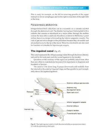
The inguinal canal (Fig. 45)
This canal represents the oblique passage taken through the lower abdomi-
nal wall by the testis and cord (the round ligament in the female).
Questions on the anatomy of this region are probably asked more often
than any other in examinations because of its importance in diagnosis and
treatment of hernias.
The canal is 1.5in (4cm) long. It passes downwards and medially from
the internal to the external inguinal rings and lies parallel to and immedi-
ately above, the inguinal ligament.
Fig. 45◊The right inguinal canal (a) with the external oblique aponeurosis intact,
(b) with the aponeurosis laid open.