
Page 82 - Clinical Anatomy
P. 82
ECA2 7/18/06 6:42 PM Page 67
Peritoneal cavity 67
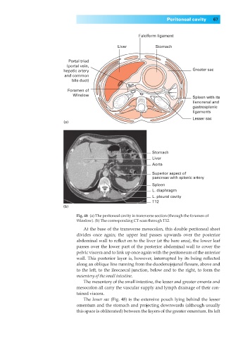
Falciform ligament
Liver Stomach
Portal triad
(portal vein,
hepatic artery Greater sac
and common
bile duct)
Foramen of
Winslow
Spleen with its
lienorenal and
gastrosplenic
ligaments
Lesser sac
(a)
Stomach
Liver
Aorta
Superior aspect of
pancreas with splenic artery
Spleen
L. diaphragm
L. pleural cavity
T12
(b)
Fig. 48◊(a) The peritoneal cavity in transverse section (through the foramen of
Winslow). (b) The corresponding CT scan through T12.
At the base of the transverse mesocolon, this double peritoneal sheet
divides once again; the upper leaf passes upwards over the posterior
abdominal wall to reflect on to the liver (at the bare area), the lower leaf
passes over the lower part of the posterior abdominal wall to cover the
pelvic viscera and to link up once again with the peritoneum of the anterior
wall. This posterior layer is, however, interrupted by its being reflected
along an oblique line running from the duodenojejunal flexure, above and
to the left, to the ileocaecal junction, below and to the right, to form the
mesentery of the small intestine.
The mesentery of the small intestine, the lesser and greater omenta and
mesocolon all carry the vascular supply and lymph drainage of their con-
tained viscera.
The lesser sac (Fig. 48) is the extensive pouch lying behind the lesser
omentum and the stomach and projecting downwards (although usually
this space is obliterated) between the layers of the greater omentum. Its left