
Page 29 - Critical Care Notes
P. 29
4223_Tab01_001-044 29/08/14 10:46 AM Page 23
23
Intra-Arterial Waveform
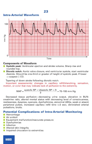
100
mm Hg
0
Time
Components of Waveform
■ Systolic peak: Ventricular ejection and stroke volume. Sharp rise and
rounded top.
■ Dicrotic notch: Aortic valve closure, end ventricular systole, start ventricular
diastole. Should be one-third or greater of height of systolic peak. If lower
→ suspect ↓ CO.
Tapering of down stroke following dicrotic notch.
Important assessments: changes in capillary refill/blanching, sensation,
motion, or color that may indicate lack of perfusion to the extremity.
systolic BP + (diastolic BP × 2)
MAP = = 70–100 mm Hg
3
Decreased tissue perfusion—decreasing urine output, elevation in BUN:
creatinine ratio, altered mental status with decreasing level of consciousness,
restlessness, dyspnea, cyanosis, dysrhythmias, abnormal ABGs, weak or absent
peripheral pulses, increased capillary refill time (>3 sec), diminished arterial
pulsations, bruits.
Potential Complications of Intra-Arterial Monitoring
■ Hemorrhage
■ Air emboli
■ Equipment malfunction/inaccurate pressure
■ Dysrhythmias
■ Infection
■ Altered skin integrity
■ Impaired circulation to extremities
BASICS