
Page 34 - Critical Care Notes
P. 34
4223_Tab01_001-044 29/08/14 10:46 AM Page 28
BASICS
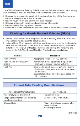
AACN Verification of Feeding Tube Placement Guidelines 2009: Use a variety
(two or more) of bedside methods to check feeding tube location:
■ Observe for a change in length of the external portion of the feeding tube.
Monitor tube position at 4-hr intervals.
■ Review routine CXR and abdominal x-ray reports.
■ Observe changes in volume and appearance of aspirate.
■ Measure pH of feeding tube aspirates.
■ Obtain an x-ray to confirm tube position when in doubt about tube’s location.
Checking for Gastric Residual Volumes (GRVs)
1. Assess GRVs every 4 hr during initial 48 hr of feeding, then 4–6 hr for con-
tinuous feeding and prior to bolus feeding.
2. Using a 30- to 60-mL syringe, withdraw gastric contents from the feeding tube.
Note volume of formula. Flush with 30 mL water. Assess for pain, abdominal
distention, “feeling full or bloated,” nausea, and emesis. The following are
based on the A.S.P.E.N. Enteral Nutrition Practice Recommendations.
Volume Indication
<250–300 mL Acceptable residual for ICU patients.
>250 mL for 2 consecutive Administer metoclopramide (Reglan) and
checks continue to assess residual volume.
>500 mL Stop feeding and reassess patient. Resume
feeding at slower rate, amount, or frequency
as indicated.
Several algorithms have been proposed for
ICU patients.
Enteral Tube Feeding Complications
Mechanical Complications Interventions
Nasopharyngeal discomfort • Reposition tube.
Esophageal ulceration or bleeding • Consider PEG or PEJ tube.
esophageal varices
Clogged tube • Flush with lukewarm water after
every feeding.
• Hospital protocol:
Continued
28