
Page 282 - Clinical Application of Mechanical Ventilation
P. 282
248 Chapter 9
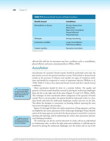
TABLE 9-4 Abnormal Breath Sounds and Related Conditions
Breath Sound Conditions
Diminished or absent Airway obstruction
Atelectasis
Main-stem intubation
Pleural effusion
Pneumothorax
Wheezes Airway narrowing
Inspiratory crackles Lung consolidation
Pulmonary edema
Coarse crackles Excessive secretions
© Cengage Learning 2014
affected side with less air movement may have conditions such as consolidation,
pleural effusion, atelectasis, and pneumothorax (White, 2003).
Auscultation
Auscultation of a patient’s breath sounds should be performed each time the
practitioner assesses the patient/ventilator system. Diminished or absent breath
sounds or the presence of wheezes and crackles are signs of ventilatory prob-
lems and should be recognized as causes of respiratory distress (Wilkins et al.
1998). Table 9-4 shows these abnormal breath sounds and their related clinical
conditions.
Chest auscultation should be done in a systemic fashion. The quality and
A side-to-side technique quantity of breath sounds should be assessed by placing the stethoscope diaphragm
of chest auscultation allows
comparison of the quantity of from the left to the right side of the chest (Figures 9-4 and 9-5) (White, 2003).
breath sounds between the
left and right lungs. This technique of chest auscultation allows comparison of the quantity of breath
sounds. Prior to the procedure, the patient should be instructed to take in a slow,
deep breath each time the stethoscope diaphragm touches and rests on the skin.
This allows the therapist to concentrate on listening without repeating the same
instruction throughout the procedure.
Figures 9-6 through 9-8 show the surface projections of lung segments, and they
are helpful for the correct placement of the stethoscope diaphragm. Proper identifi-
cation of the lung segments involved in the disease process is essential for consistent
charting and reporting, and for performing the correct chest percussion and pos-
A cuff leak may be pres- tural drainage procedures.
ent if distinct air movement The stethoscope can also be used for detection of a leaky cuff on an endotracheal
can be heard toward the end
of a mechanical breath. or tracheostomy tube, as well as for right main-stem intubation. A cuff leak may be
detected by placing the stethoscope diaphragm over the trachea and on top of the
Copyright 2013 Cengage Learning. All Rights Reserved. May not be copied, scanned, or duplicated, in whole or in part. Due to electronic rights, some third party content may be suppressed from the eBook and/or eChapter(s).
Editorial review has deemed that any suppressed content does not materially affect the overall learning experience. Cengage Learning reserves the right to remove additional content at any time if subsequent rights restrictions require it.