
Page 286 - Clinical Application of Mechanical Ventilation
P. 286
252 Chapter 9
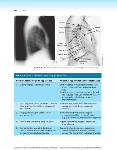
Lung apex
Trachea Sternum
Aorta
Scapula
Posterior ribs
(superimposed)
Hilar region
Heart shadow
© Cengage Learning 2014 vertebrae Costophrenic © Cengage Learning 2014
Thoracic
Diaphragm
angle
Figure 9-10 A normal lateral chest radiograph and corresponding anatomical structures.
TABLE 9-5 Normal and Abnormal Chest Radiograph Appearance
Normal Chest Radiograph Appearance Abnormal Appearance and Probable Cause
1. Midline trachea and mediasternum Shift of trachea or mediasternum may occur
due to patient rotation during taking of
X-ray.
Shift of trachea or mediasternum to affected
side (e.g., atelectasis, pulmonary fibrosis) or
to the unaffected side (e.g., tension
pneumonthorax)
2. Dark lung parenchyma with mild scattered Infiltrates (large amount of white shadows)
white shadows (normal lung tissues and suggest accumulation of secretions,
small vessels) atelectasis.
3. Sharply pointed right and left costo- Blunted costophrenic angle suggests
phrenic angels accumulation of fluid in pleural space
(e.g., pleural effusion, hemothorax, empyema)
4. Smooth and continuing bony structures Jagged appearance suggests fracture or
broken bones.
5. Cardiac shadow along the diaphragm Congestive heart may cause the cardiac
line is , 50% of the distance between the shadow to exceed 50% of the distance
right and left costophrenic angles. between the right and left costophrenic angels.
© Cengage Learning 2014
Copyright 2013 Cengage Learning. All Rights Reserved. May not be copied, scanned, or duplicated, in whole or in part. Due to electronic rights, some third party content may be suppressed from the eBook and/or eChapter(s).
Editorial review has deemed that any suppressed content does not materially affect the overall learning experience. Cengage Learning reserves the right to remove additional content at any time if subsequent rights restrictions require it.