
Page 285 - Clinical Application of Mechanical Ventilation
P. 285
Monitoring in Mechanical Ventilation 251
Imaging
The chest radiograph is the most common method to evaluate the conditions
of the thoracic structure, lungs, pleural space, inserted catheters, lines, and
tubes. Interpretation of the chest radiograph is beyond the scope of this sec-
tion. Readers are encouraged to use additional resources on the fundamentals
and clinical application of chest radiography. This section reviews the normal
posterior-anterior (PA) and lateral chest radiographs, the major anatomical
structures on the chest radiograph, and the primary reason for using a lateral
chest radiograph.
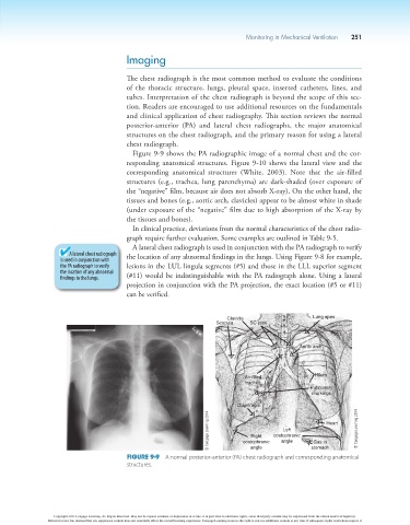
Figure 9-9 shows the PA radiographic image of a normal chest and the cor-
responding anatomical structures. Figure 9-10 shows the lateral view and the
corresponding anatomical structures (White, 2003). Note that the air-filled
structures (e.g., trachea, lung parenchyma) are dark-shaded (over exposure of
the “negative” film, because air does not absorb X-ray). On the other hand, the
tissues and bones (e.g., aortic arch, clavicles) appear to be almost white in shade
(under exposure of the “negative” film due to high absorption of the X-ray by
the tissues and bones).
In clinical practice, deviations from the normal characteristics of the chest radio-
graph require further evaluation. Some examples are outlined in Table 9-5.
A lateral chest radiograph is used in conjunction with the PA radiograph to verify
A lateral chest radiograph the location of any abnormal findings in the lungs. Using Figure 9-8 for example,
is used in conjunction with
the PA radiograph to verify lesions in the LUL lingula segments (#5) and those in the LLL superior segment
the location of any abnormal
findings in the lungs. (#11) would be indistinguishable with the PA radiograph alone. Using a lateral
projection in conjunction with the PA projection, the exact location (#5 or #11)
can be verified.
Clavicle Lung apex
Scapula SC joint
Aortic arch
Air-filled Hilum
trachea
Pulmonary
markings
Diaphragm
© Cengage Learning 2014 costophrenic costophrenic Gas in Heart © Cengage Learning 2014
Left
Right
angle
stomach
angle
Figure 9-9 A normal posterior-anterior (PA) chest radiograph and corresponding anatomical
structures.
Copyright 2013 Cengage Learning. All Rights Reserved. May not be copied, scanned, or duplicated, in whole or in part. Due to electronic rights, some third party content may be suppressed from the eBook and/or eChapter(s).
Editorial review has deemed that any suppressed content does not materially affect the overall learning experience. Cengage Learning reserves the right to remove additional content at any time if subsequent rights restrictions require it.