
Page 410 - Clinical Application of Mechanical Ventilation
P. 410
376 Chapter 12
STRATEGIES TO IMPROVE VENTILATION
Hypoventilation causes respiratory acidosis (ventilatory failure) and hypoxemia
PaCO 2 .45 mm Hg is if supplemental oxygen is not provided to the patient. The best measure of
indicative of hypoventilation
(the normal PaCO 2 for COPD a patient’s ventilatory status is the PaCO level. The normal PaCO is 35 to
2
2
patients is about 50 mm Hg). 45 mm Hg; PaCO greater than 45 mm Hg is indicative of hypoventilation.
2
For COPD patients, however, the acceptable PaCO should be the patient’s
2
normal value upon last hospital discharge, and generally it is about 50 mm Hg.
When the PaCO level goes above this value, significant hypoventilation may
2
be present.
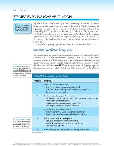
Strategies for improving a patient’s ventilation are summarized in Table 12-2.
Increase Ventilator Frequency
The most common approach to improve minute ventilation is to increase the ventila-
tor frequency (f ). This may be the control frequency in assist/control, the mandatory
frequency in synchronized intermittent mandatory ventilation, or other modes of ven-
tilation that regulate the frequency of the ventilator. However, the ventilator frequency
should not exceed 20/min as auto-PEEP may occur at or above this frequency, especially
auto-PEEP: Unintentional PEEP
associated with pressure support during pressure support ventilation (MacIntyre, 1986; Shapiro, 1994). The following
ventilation, high tidal volume and
frequency, inadequate inspiratory
flow, excessive I-time, inadequate
E-time, and air trapping. TABLE 12-2 Strategies to Improve Ventilation
Priority Methods
1 Increase ventilator frequency
Control frequency in assist/control mode
Intermittent mandatory ventilation (IMV) frequency
Synchronized IMV frequency
2 Increase spontaneous tidal volume
Nutritional support and reconditioning of respiratory muscles
Administer bronchodilators
Initiate pressure support ventilation (PSV)
Use largest endotracheal tube possible
3 Increase ventilator tidal volume
Tidal volume in volume-controlled ventilation.
Pressure in pressure-controlled ventilation.
4 Reduce mechanical deadspace
mechanical deadspace: Volume
of gas contained in the equipment Use low-compliance ventilator circuit
and supplies (e.g., endotracheal Cut endotracheal tube to appropriate length
tube, ventilator circuit) that does Perform tracheotomy
not take part in gas exchange.
5 Consider high frequency jet or oscillatory ventilation
© Cengage Learning 2014
Copyright 2013 Cengage Learning. All Rights Reserved. May not be copied, scanned, or duplicated, in whole or in part. Due to electronic rights, some third party content may be suppressed from the eBook and/or eChapter(s).
Editorial review has deemed that any suppressed content does not materially affect the overall learning experience. Cengage Learning reserves the right to remove additional content at any time if subsequent rights restrictions require it.