Page 711 - Clinical Application of Mechanical Ventilation
P. 711

Case	Studies  677


                                             was	slow,	indicating	poor	peripheral	perfusion,	but	the	extremities	were	warm.
                                             The	anterior	fontanel	was	soft	with	slightly	overlapping	sutures.	Breath	sounds	were
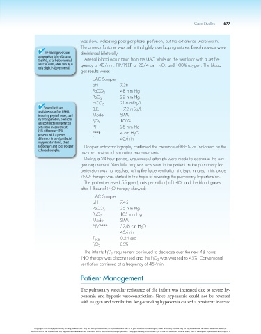
                            The blood gases show   diminished	bilaterally.
                          oxygenation failure because
                          the PaO 2  is far below normal   Arterial	blood	was	drawn	from	the	UAC	while	on	the	ventilator	with	a	set	fre-
                          and the PaCO 2  of 48 mm Hg is   quency	of	40/min,	PIP/PEEP	of	28/4	cm	H O,	and	100%	oxygen.	The	blood
                          only slightly above normal.                             2
                                             gas	results	were:
                                             	     UAC	Sample
                                             	     pH	          7.28
                                             	     PaCO 2	      48	mm	Hg
                                             	     PaO 2	       22	mm	Hg
                                                        -
                                             	     HCO 	        21.6	mEq/L
                                                        3
                            Several tests are   	  B.E.	        27.2	mEq/L
                          available to confirm PPHN,
                          including physical exam, labil-  	  Mode	  SIMV
                          ity of oxygenation, preductal   	  FO  100%
                          and postductal oxygenation   I  2
                          saturation measurements   	  PIP	     28	mm	Hg
                          (5% difference2PDA   	   PEEP	        4	cm	H O
                          presents with a greater                     2
                          difference in pre-/postductal   	  f	  40/min
                          oxygen saturations), chest
                          radiograph, and color Doppler   Doppler	echocardiography	confirmed	the	presence	of	PPHN	as	indicated	by	the
                          echocardiography.
                                             pre-	and	postductal	saturation	measurements.
                                               During	a	24-hour	period,	unsuccessful	attempts	were	made	to	decrease	the	oxy-
                                             gen	requirement.	Very	little	progress	was	seen	in	the	patient	as	the	pulmonary	hy-
                                             pertension	was	not	resolved	using	the	hyperventilation	strategy.	Inhaled	nitric	oxide
                                             (iNO)	therapy	was	started	in	the	hope	of	reversing	the	pulmonary	hypertension.
                                               The	patient	received	55	ppm	(parts	per	million)	of	iNO,	and	the	blood	gases
                                             after	1	hour	of	iNO	therapy	showed:
                                             	     UAC	Sample
                                             	     pH	          7.45
                                             	     PaCO 2	      35	mm	Hg
                                             	     PaO 2	       105	mm	Hg
                                             	     Mode	        SIMV
                                             	     PIP/PEEP	    32/6	cm	H O
                                                                          2
                                             	     f	           45/min
                                             	     T INSP	      0.24	sec
                                             	     FO 2	        85%
                                                    I
                                               The	infant’s	FO 	requirement	continued	to	decrease	over	the	next	48	hours.
                                                            2
                                                          I
                                             iNO	therapy	was	discontinued	and	the	FO 	was	weaned	to	45%.	Conventional
                                                                                 2
                                                                               I
                                             ventilation	continued	at	a	frequency	of	45/min.
                                             Patient Management


                                             The pulmonary vascular resistance of the infant was increased due to severe hy-
                                             poxemia  and  hypoxic  vasoconstriction.  Since  hypoxemia  could  not  be  reversed
                                             with oxygen and ventilation, long-standing hypoxemia caused a persistent increase


                        Copyright 2013 Cengage Learning. All Rights Reserved. May not be copied, scanned, or duplicated, in whole or in part. Due to electronic rights, some third party content may be suppressed from the eBook and/or eChapter(s).
                      Editorial review has deemed that any suppressed content does not materially affect the overall learning experience. Cengage Learning reserves the right to remove additional content at any time if subsequent rights restrictions require it.
   706   707   708   709   710   711   712   713   714   715   716