
Page 137 - Cardiac Nursing
P. 137
9/0
0
009
9/2
1-1
p11
qxd
31.
8:2
13
e 1
ara
Apt
M
4 A
g
P
K34
LWB
05_
0-c
LWBK340-c05_p111-131.qxd 09/09/2009 08:24 AM Page 113 Aptara
L L LWB K34 0-c 05_ p11 1-1 31. qxd 0 9/0 9/2 009 0 0 8:2 4 A M P a a g e 1 13 Apt ara
C HAPTER 5 / Atherosclerosis, Inflammation, and Acute Coronary Syndrome 113
mortality. However, the underlying complexity of the disease has and histochemically (by staining for lipid deposits) in the intima.
34
made precise delineation of the cellular and molecular mechanisms Type I lesions are most often observed in infants and children, al-
involved difficult. Over the past decade, new investigative tools have though they are readily identifiable in adults with little atheroscle-
contributed to a clearer picture of the molecular mechanisms under- rosis or in areas of the vasculature not prone to arteriosclerosis.
lying the development of atherosclerotic plaque. It is clear that ath- These lesions occur in regions of the intima that display adaptive in-
erosclerosis is not simply an inevitable consequence of ageing. timal thickening caused by the hemodynamic force of blood flow.
These regions eventually evolve into types II and III lesions. Al-
American Heart Association Lesion though more common in early adulthood, the occurrence of type
34,35
Classification System III lesions has been reported as early as the first year of life. The
accumulation of intimal foam cells is a consequence and a marker
More than a decade ago, the American Heart Association (AHA) of pathological accumulation of atherogenic lipoproteins.
endeavored to provide an organized system for the categorization
of lesions based on histological and morphological data. 28 This Type II Lesions
system has helped standardize research in atherosclerosis, though Type II lesions, also known as fatty streaks that are visible on gross
modifications have been proposed. 29 inspection, are yellow spots or streaks on arterial intima. The trans-
Coronary artery lesions can be grouped into seven major types migration of macrophages into the subendothelial space and their
(I to VII). 28,30–33 Consistent morphologic data would seem to in- subsequent transformation into foam cells produces an adaptive in-
dicate that each lesion type is relatively stable and will not progress timal thickening, which may obscure the fatty streak, potentially
to the next lesion type without additional factors or pressures. leading to an underestimate of the extent of these lesions. Recruit-
While the advanced lesions (types IV to VII) can manifest clini- ment of macrophages to the intima marks one of the defining
cally, the early lesions (types I to III) are clinically silent and can events in the initiation of the atherosclerotic lesion. Specific adhe-
be organized temporally. Types I and II are generally found in sion molecules expressed on the surface of vascular endothelial cells
children, whereas type III tends to occur later and bridges early mediate leucocyte adhesion. In addition, modified lipoproteins
and advanced lesions. Perhaps the most important observation is contain oxidized phospholipids that induce the expression of adhe-
that the clinically silent lesions (types I to III) have been shown sion molecules and cytokines implicated in early atherogenesis. 22
capable of regression in animal models. Advanced lesions are gen- Progression of atheroma involves accumulation of smooth mus-
erally disorganized and lead to thickening and eventual compro- cle cells that elaborate extracellular matrix macromolecules. Micro-
mise of the vessel wall. Lipid-laden macrophages (termed “foam scopic examination of type II lesions reveals that the foam cells are
cells”) are the predominant cellular components of type I lesions. more organized, stratifying into layers, and that smooth muscle cells
In types II and III lesions, intimal smooth muscle cells dominate, also begin to show signs of intracellular lipid accumulation. The
with minimal involvement of lymphocytes, plasma cells, and mast properties of these lesions results in continued recruitment of
cells in the pathological processes. This group of inflammatory macrophage and evidence suggests that T-lymphocytes and mast
cells becomes quite active in advanced (types IV and V) lesions. cells (components of the immune system) begin to invade the le-
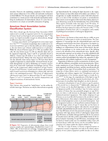
Figures 5-2 and 5-3 summarize the essential characteristics and sion. 30,36,37 At this stage, the preponderance of the lipid in type II
temporal occurrence of atherosclerotic lesions. lesions resides in cells, with the majority found in foam cells. A lim-
ited amount of extracellular lipid (droplets) can also be detected.
Type I Lesions Consistent colocalization of type II lesions to specific portions
Type I lesions, often termed the “initial lesion,” are the earliest de- of the arterial tree is characteristic. 38 Additionally, subgroups of
tectable lesion type. The lesion can only be observed microscopically type II lesions can be described dependent on their location and
the lipoprotein profile of the individual. Type IIa lesions represent
the subset of lesions that may potentially progress to type III le-
sions over time or with increases in atherogenic (triglyceride- and
Complicated
Foam Fatty Intermediate Fibrous lesion or cholesterol-enriched) lipoproteins. This smaller subgroup of type
cells streak lesion Atheroma plaque rupture II lesions occurs in predictable locations in the arterial tree (proxi-
mal to bifurcations), where adaptive intimal thickenings occur, and
are also termed progression-prone or advanced lesion-prone. Type
IIb (progression-resistant or advanced lesion-resistant) lesions con-
sist of the larger subset of type II lesions that are less likely to
progress and are located in regions with relatively normal intima
with little subendothelial smooth muscle cell invasion or prolifera-
tion. Type IIb lesions do have the potential to progress, particularly
in persons with high plasma levels of atherogenic lipoproteins.
From first decade From third decade From fourth decade Type IIb lesions are further distinguished from IIa by the presence
Smooth of smooth muscle cells that produce intercellular matrix in the re-
muscle Thrombosis, gion of adaptive thickening. In type IIb lesions, macrophages with-
Growth mainly by lipid accumulation
and haematoma out lipid are found mostly near the endothelial surface, foam cells
collagen
are found deeper within the intima, and the extracellular lipid ac-
■ Figure 5-2 Progression of atheromatous plaque from initial cumulates even deeper within the adaptive thickening.
lesion to complex and ruptured plaque. (Modified from Grech, E. D. The fate of a type II lesion, to become progression-prone or
[2003]. ABC of interventional cardiology: Pathophysiology and in- progression-resistant, is dependent not only on the relative athero-
vestigation of coronary artery disease. BMJ, 326[7397], 1027–1030.)6 genicity of one’s plasma lipoprotein profile but also on the direct
6