
Page 140 - Cardiac Nursing
P. 140
LWBK340-c05_p111-131.qxd 09/09/2009 08:24 AM Page 116 Aptara
116 PA R T II / Physiologic and Pathologic Responses
changes affect the vulnerability of the plaque and propensity for
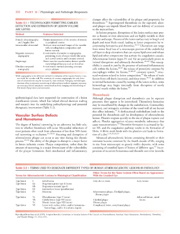
Table 5-1 ■ TECHNOLOGIES PERMITTING EARLIER thrombosis. 10 Superimposed thrombosis on the ruptured, ulcer-
DETECTION AND ESTIMATION OF LESION VOLUME ated plaque can impede blood flow and the delivery of nutrients
ARE LISTED to the myocardium.
As lesions progress, disruptions of the lesion surface may pres-
Method Features Detected
ent as fissures or even ulcerations and are highly variable in their
B-mode ultrasonography Permits measurement of the severity of stenosis severity and scope. Fissures of the lesion surface vary in length and
and Doppler flow in peripheral arteries depth and most likely reseal, leading to lesion progression by in-
Intravascular ultrasound Produces cross-sectional images of the vascular corporating hematoma and thrombus. 42,43 Ulcerations can range
wall, revealing lesion composition and
lumen contour from minor focal loss of a microscopic portion of the endothelial
Magnetic resonance A noninvasive alternative to angiography, cell layer to deep ulcerations that can expose lipid cores and release
angiography permitting study of major vessels (the aorta lipid and other components that activate the coagulation cascade.
and carotid arteries and coronary arteries) Atheromatous lesions (types IV and Va) are particularly prone to
Angioscopy Direct vascular vascularization detects specific intimal disruptions and ultimately thrombosis. 42–45 This suscep-
morphological features such as thrombus
Ultrafast computed A noninvasive method detecting coronary tibility is caused in part by the presence of activated inflammatory
tomography artery calcium cells within the lesions, 46,47 the release of proteolytic enzymes by
macrophages within the lesions, 42,48,49 coronary spasm, 50 struc-
45
While angiography is the definitive method for evaluation of the vascular lumen, it can- tural weakness related to lesion composition, the release of toxic
not detail the vascular wall. The sensitivity of coronary angiography for early detec- factors from cell death (necrosis), and shear stress. 39,51 In addition
tion of atherosclerosis may be increased by these methods. Emerging methodologies
that may allow noninvasive monitoring of atherosclerosis include magnetic resonance to intimal hematoma caused by tearing of the lesion surface, some
spectroscopy, labeled antiplatelet monoclonal antibody imaging and radiolabeling of hemorrhage may begin internally from disruption of newly
low-density lipoproteins and monocytes. formed vessels within the lesion. 52
Thrombosis
epidemiological data have supported the construction of a lesion Although plaque disruption and thrombosis can be separate
classification system, which has helped clinical decision making processes, they appear to be interrelated. Thrombosis formation
and research into the underlying pathophysiology and potential may be exacerbated by changes in the endothelium. Contractility,
therapeutic intervention (Table 5-2). secretory, and mitogenic activities of the vessel wall all are factors
that affect ischemia. 10 A dysfunctional endothelium leads to the
Vascular Surface Defects potential for thrombosis and the development of atherosclerotic
and Hematoma lesions. Platelets migrate quickly to the site of plaque rupture and
adhere. Platelet aggregation releases metabolic substances that
53
The degree of luminal narrowing by an atheroma has little rela- cause vasoconstriction. Thrombin formation is activated by fac-
tion to whether thrombosis will occur. Myocardial infarctions of tor XII, and the coagulation pathway results in the formation of
most patients often result from atheromas of less than 50% lumi- fibrin. A fibrin mesh binds with the platelets and leads to forma-
nal narrowing or occlusion. 11,18,41 Fissuring and disruption of tion of a clot. 3,11,25,26,54
atherosclerotic plaque can occur at any time during this chronic Advanced atherosclerotic lesions containing thrombi or their
process. 12,14 The ability of the plaque to disrupt is a major factor remnants become common by the fourth decade of life, ranging
in future ischemic events. Plaque composition, rather than the in size from microscopic to grossly visible deposits, with some
amount of narrowing, is a major determinant of the vulnerability consisting of stratified layers of lesions of different ages. 55 Incor-
of the plaque formation. Both mechanical and inflammatory poration of recurrent hematomas and thrombi over time (months
Table 5-2 ■ TERMS USED TO DESIGNATE DIFFERENT TYPES OF HUMAN ATHEROSCLEROTIC LESIONS IN PATHOLOGY
Other Terms for the Same Lesions Often Based on Appearance
Terms for Atherosclerotic Lesions in Histological Classification With the Unaided Eye
Type lesion I Initial lesion Fatty dot or streak Early lesions
Type lesion IIa Progression-prone type II lesion
IIb Progression-resistant type II
Type lesion III Intermediate lesion (preatheroma)
Type lesion IV Atheroma Atheromatous plaque, fibrolipid plaque,
fibrous plaque
Type lesion Va Fibroatheroma (type V lesion) Advanced lesions, raised
Vb Calcific lesion (type VII lesion) Calcified plaque lesions
Vc Fibrotic lesion (type VIII lesion) Fibrous plaque
Type lesion VI Lesion with surface defect, and/or hematoma– Complicated lesion, complicated plaque
hemorrhage, and/or thrombotic deposit
Reproduced from Stary et al. (1995). A report from the Committee on Vascular Lesions of the Council on Arteriosclerosis, American Heart Association. Arteriosclerosis, Thrombosis,
and Vascular Biology, 15, 1512–1531.