Page 138 - Cardiac Nursing
P. 138

009
                                    009
                                  9/2
                                  9/2
                                        8:2
                                        8:2
                                        0
                                        0
                                9/0
                         31.
                           qxd
                       1-1
                         31.
                               0
                                9/0
                           qxd
                               0
                                           4 A
                                                    14
                                                    14
                                                  e 1
                                                  e 1
                                                         ara
                                                         ara
                                                       Apt
                                                       Apt
                                                 g
                                             M
                                               P
                                           4 A
                                             M
                                                a
                                                 g
                                               P
                                                a
               0-c
               0-c
            K34
                 05_
         L L LWB
                 05_
                    p11
                    p11
                       1-1
            K34
         LWB
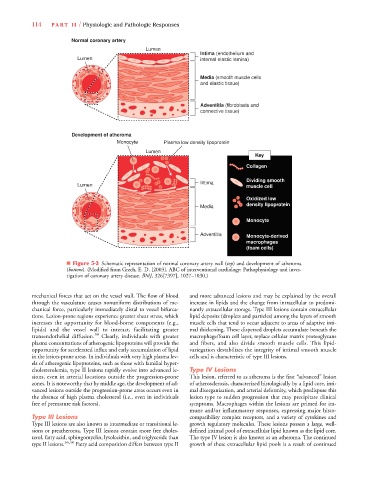
         LWBK340-c05_p111-131.qxd  09/09/2009  08:24 AM  Page 114 Aptara
                  114    PA R T  I I / Physiologic and Pathologic Responses
                               Normal coronary artery
                                                        Lumen
                                                                         Intima (endothelium and
                                 Lumen                                   internal elastic lamina)
                                                                         Media (smooth muscle cells
                                                                         and elastic tissue)
                                                                         Adventitia (fibroblasts and
                                                                         connective tissue)
                               Development of atheroma
                                              Monocyte       Plasma low density lipoprotein
                                                        Lumen
                                                                                           Key
                                                                                         Collagen
                                                                                         Dividing smooth
                                                                         Intima
                                 Lumen                                                   muscle cell
                                                                                         Oxidized low
                                                                                         density lipoprotein
                                                                         Media
                                                                                         Monocyte
                                                                         Adventitia      Monocyte-derived
                                                                                         macrophages
                                                                                         (foam cells)
                              ■ Figure 5-3 Schematic representation of normal coronary artery wall (top) and development of atheroma
                              (bottom). (Modified from Grech, E. D. [2003]. ABC of interventional cardiology: Pathophysiology and inves-
                              tigation of coronary artery disease. BMJ, 326[7397], 1027–1030.)
                  mechanical forces that act on the vessel wall. The flow of blood  and more advanced lesions and may be explained by the overall
                  through the vasculature causes nonuniform distributions of me-  increase in lipids and the change from intracellular to predomi-
                  chanical force, particularly immediately distal to vessel bifurca-  nantly extracellular storage. Type III lesions contain extracellular
                  tions. Lesion-prone regions experience greater shear stress, which  lipid deposits (droplets and particles) among the layers of smooth
                  increases the opportunity  for  blood-borne components (e.g.,  muscle cells that tend to occur adjacent to areas of adaptive inti-
                  lipids) and the vessel wall to interact,  facilitating greater  mal thickening. These dispersed droplets accumulate beneath the
                  transendothelial  diffusion. 39  Clearly, individuals with greater  macrophage/foam cell layer, replace cellular matrix proteoglycans
                  plasma concentrations of atherogenic lipoproteins will provide the  and  fibers, and also  divide smooth muscle cells. This  lipid-
                  opportunity for accelerated influx and early accumulation of lipid  variegation destabilizes the integrity of intimal smooth muscle
                  in the lesion-prone areas. In individuals with very high plasma lev-  cells and is characteristic of type III lesions.
                  els of atherogenic lipoproteins, such as those with familial hyper-
                  cholesterolemia, type II lesions rapidly evolve into advanced le-  Type IV Lesions
                  sions, even in arterial locations outside the progression-prone  This lesion, referred to as atheroma is the first “advanced” lesion
                  zones. It is noteworthy that by middle age, the development of ad-  of atherosclerosis, characterized histologically by a lipid core, inti-
                  vanced lesions outside the progression-prone areas occurs even in  mal disorganization, and arterial deformity, which predispose this
                  the absence of high plasma cholesterol (i.e., even in individuals  lesion type to sudden progression that may precipitate clinical
                  free of premature risk factors).                    symptoms. Macrophages within the lesions are primed for im-
                                                                      mune and/or inflammatory responses, expressing major histo-
                  Type III Lesions                                    compatibility complex receptors, and a variety of cytokines and
                  Type III lesions are also known as intermediate or transitional le-  growth regulatory molecules. These lesions possess a large, well-
                  sions or preatheroma. Type III lesions contain more free choles-  defined intimal pool of extracellular lipid known as the lipid core.
                  terol, fatty acid, sphingomyelin, lysolecithin, and triglyceride than  The type IV lesion is also known as an atheroma. The continued
                  type II lesions. 36,40  Fatty acid composition differs between type II  growth of these extracellular lipid pools is a result of continued
   133   134   135   136   137   138   139   140   141   142   143