
Page 26 - Cardiac Nursing
P. 26
92806_c01.qxd 11/21/11 10:30 AM Page 2
2 PA R T I / Anatomy and Physiology
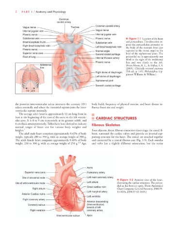
■ Figure 1-1 Location of the heart
and pericardium. This dissection ex-
poses the pericardialsac posterior to
the body of the sternum from just
superior to the sterna angel to the
level of the xiphisternal joint. The
pericardial sac is approximately one
third to the right of the midsternal
line and two thirds to the left.
(From Moore, K. L., & Dalley, A. F.
[2005]. Clinically oriented anatomy
[5th ed., p. 145]. Philadelphia: Lip-
pincott Williams & Wilkins.)
the posterior interventricular sulcus intersects the coronary (AV) body build, frequency of physical exercise, and heart disease in-
sulcus externally and where the interatrial septum joins the inter- fluence heart size and weight.
ventricular septum internally.
The average adult heart is approximately 12 cm long from its
base at the beginning of the root of the aorta to the left ventric- CARDIAC STRUCTURES
ular apex. It is 8 to 9 cm transversely at its greatest width, and
6 cm thick anteroposteriorly. Tables have been derived to indicate Fibrous Skeleton
normal ranges of heart size for various body weights and
heights. 1 Four adjacent, dense, fibrous connective tissue rings, the annuli fi-
The adult male heart comprises approximately 0.43% of body brosi, surround the cardiac valves and provide an internal sup-
weight, typically 280 to 350 g, with an average weight of 300 g. porting structure for the heart. The annuli are attached together
The adult female heart comprises approximately 0.40% of body and connected by a central fibrous core (Fig. 1-5). Each annulus
weight, 230 to 300 g, with an average weight of 250 g. 2,3 Age, and valve has a slightly different orientation, but the entire
■ Figure 1-2 Anterior view of the heart,
illustrating the cardiac structures. The pericar-
dial sac has been cut open. (From Anatomical
Chart Company, General Anatomy, 2008-05-
14 0614, 2008-07-13 1449.)