
Page 28 - Cardiac Nursing
P. 28
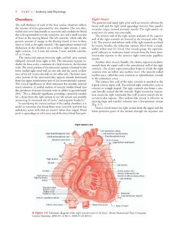
92806_c01.qxd  11/21/11  10:30 AM  Page 4


                     4   PA R T  I / Anatomy and Physiology

                     Chambers                                           Right Heart
                                                                        The posterior and septal right atrial walls are smooth, whereas the
                     The wall thickness of each of the four cardiac chambers reflects
                                                                        lateral wall and the right atrial appendage (auricle) have parallel
                     the amount of force generated by that chamber. The two thin-
                                                                        muscular ridges, termed pectinate muscles. The right auricle ex-
                     walled atria serve functionally as reservoirs and conduits for blood
                                                                        tends over the aortic root externally.
                     that is being funneled into the ventricles; they add a small amount
                                                                           The inferior wall of the right atrium and part of the superior
                     of force to the moving blood. The left ventricle, which adds the
                                                                        wall of the right ventricle are formed by the tricuspid valve (Fig.
                     greatest amount of energy to the flowing blood, is two to three
                                                                        1-6). The anterior and inferior walls of the right ventricle are lined
                     times as thick as the right ventricle. The approximate normal wall
                                                                        by muscle bundles, the trabeculae carneae, which form a rough-
                     thicknesses of the chambers are as follows: right atrium, 2 mm;
                                                                        walled inflow tract for blood. One muscle group, the septomar-
                     right ventricle, 3 to 5 mm; left atrium, 3 mm; and left ventricle,
                                                                        ginal trabecula or moderator band, extends from the lower inter-
                     13 to 15 mm.
                                                                        ventricular septum to the anterior right ventricular papillary
                       The interatrial septum between right and left atria extends
                                                                        muscle.
                     obliquely forward from right to left. The interatrial septum in-
                                                                           Another thick muscle bundle, the christa supraventricularis,
                     cludes the fossa ovalis, a remnant of a fetal structure, the foramen
                                                                        extends from the septal wall to the anterolateral wall of the right
                     ovale. The lower portion of the interatrial septum is formed by the
                                                                        ventricle. The christa supraventricularis helps to divide the right
                     lower medial right atrial wall on one side and the aortic outflow
                                                                        ventricle into an inflow and outflow tract. The smooth-walled
                     tract of the left ventricular wall on the other side. The lower mus-
                                                                        outflow tract, called the conus arteriosus or infundibulum, extends
                     cular portion of the interventricular septum extends downward
                                                                        to the pulmonary artery.
                     from the upper membranous part of the interventricular septum.
                                                                           The concave free wall of the right ventricle is attached to the
                     The clinical significance of these structures has recently received
                                                                        slightly convex septal wall. The internal right ventricular cavity is
                     much attention. A pooled analysis of autopsy studies found that
                                                                        crescent or triangle shaped. The right ventricle also forms a cres-
                     the prevalence of patent foramen ovale in adults is approximately  cent laterally around the left ventricle. Right ventricular contrac-
                        4
                     26%. This is clinically significant, providing a potential conduit
                                                                        tion causes the right ventricular free wall to move toward the in-
                     for a shunt from the right atrium to the left atrium and possibly  terventricular septum. This bellows-like action is effective in
                                                 5
                     accounting for increased risk of stroke and migraine headache. 6
                                                                        ejecting large and variable volumes into a low-pressure system
                       In considering the internal surfaces of the cardiac chambers, it is
                                                                        (Fig. 1-7).
                    useful to remember that blood flows more smoothly and with less
                                                                           Venous blood enters the right atrium from the upper and the
                    turbulence across walls that are smooth rather than ridged. Blood
                                                                        lower posterior parts of the atrium through the superior and
                    pools in appendages or other areas out of the direct blood flow path.


                                ■ Figure 1-6 Schematic diagram of the right interior view of the heart. (From Anatomical Chart Company,
                                General Anatomy, 2008-05-14 0614, 2008-07-16 2010.)