
Page 31 - Cardiac Nursing
P. 31
92806_c01.qxd 11/21/11 10:30 AM Page 7
CHAPTER 1 / Cardiac Anatomy and Physiology 7
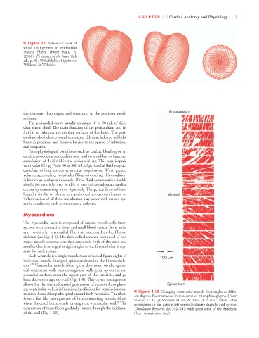
■ Figure 1-9 Schematic view of
spiral arrangement of ventricular
muscle fibers. (From Katz, A.
[2006]. Physiology of the heart [4th
ed., p. 8]. Philadelphia: Lippincott
Williams & Wilkins.)
the sternum, diaphragm, and structures in the posterior medi-
astinum.
The pericardial cavity usually contains 10 to 30 mL of thin,
clear serous fluid. The main function of the pericardium and its
fluid is to lubricate the moving surfaces of the heart. The peri-
cardium also helps to retard ventricular dilation, helps to hold the
heart in position, and forms a barrier to the spread of infections
and neoplasia.
Pathophysiological conditions such as cardiac bleeding or an
exudate-producing pericarditis may lead to a sudden or large ac-
cumulation of fluid within the pericardial sac. This may impede
ventricular filling. From 50 to 300 mL of pericardial fluid may ac-
cumulate without serious ventricular impairment. When greater
volumes accumulate, ventricular filling is impaired; this condition
is known as cardiac tamponade. If the fluid accumulation builds
slowly, the ventricles may be able to maintain an adequate cardiac
output by contracting more vigorously. The pericardium is histo-
logically similar to pleural and peritoneal serous membranes, so
inflammation of all three membranes may occur with certain sys-
temic conditions such as rheumatoid arthritis.
Myocardium
The myocardial layer is composed of cardiac muscle cells inter-
spersed with connective tissue and small blood vessels. Some atrial
and ventricular myocardial fibers are anchored to the fibrous
skeleton (see Fig. 1-5). The thin-walled atria are composed of two
major muscle systems: one that surrounds both of the atria and
another that is arranged at right angles to the first and that is sep-
arate for each atrium.
Each ventricle is a single muscle mass of nested figure eights of
individual muscle fiber path spirals anchored to the fibrous skele-
ton. 7,8 Ventricular muscle fibers spiral downward on the epicar-
dial ventricular wall, pass through the wall, spiral up on the en-
docardial surface, cross the upper part of the ventricle, and go
back down through the wall (Fig. 1-9). This vortex arrangement
allows for the circumferential generation of tension throughout
the ventricular wall; it is functionally efficient for ventricular con-
■ Figure 1-10 Changing ventricular muscle fiber angles at differ-
traction. Some fiber paths spiral around both ventricles. The fibers
ent depths. Reconstructed from a series of microphotographs. (From
form a fan-like arrangement of interconnecting muscle fibers Streeter, D. D., Jr, Spotnitz, H. M., & Patel, D. P., et al. [1969]. Fiber
8
when dissected horizontally through the ventricular wall. The orientation in the canine left ventricle during diastole and systole.
orientation of these fibers gradually rotates through the thickness Circulation Research, 24, 342–347, with permission of the American
of the wall (Fig. 1-10). Heart Association, Inc.)