
Page 29 - Cardiac Nursing
P. 29
92806_c01.qxd 11/21/11 10:30 AM Page 5
CHAPTER 1 / Cardiac Anatomy and Physiology 5
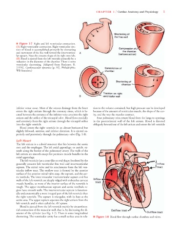
■ Figure 1-7 Right and left ventricular contraction.
(A) Right ventricular contraction. Right ventricular ejec-
tion of blood is accomplished primarily by shortening
and movement of the free wall toward the interventricu-
lar septum. Note the crescent shape of the right ventricle.
(B) Blood is ejected from the left ventricle primarily by a
reduction in the diameter of the chamber. There is some
ventricular shortening. (Adapted from Rushmer, R.
[1976]. Cardiovascular dynamics [p. 92]. Philadelphia:
WB Saunders.)
inferior venae cavae. Most of the venous drainage from the heart tion to the volume contained, but high pressure can be developed
enters the right atrium through the coronary sinus, which is lo- because of the amount of ventricular muscle, the shape of the cav-
cated between the entrance of the inferior vena cava into the right ity, and the way the muscles contract.
atrium and the orifice of the tricuspid valve. Blood flows medially Four pulmonary veins return blood from the lungs to openings
and anteriorly from the right atrium through the tricuspid orifice in the posterolateral wall of the left atrium. Blood is directed
into the right ventricle. obliquely forward out of the left atrium and enters the left ventricle
Blood enters the right ventricle in an almost horizontal but
slightly leftward, anterior, and inferior direction. It is ejected su-
periorly and posteriorly through the pulmonary valve (Fig. 1-8).
Left Heart
The left atrium is a cuboid structure that lies between the aortic
root and the esophagus. The left atrial appendage, or auricle, ex-
tends along the border of the pulmonary artery. The walls of the
left atrium are smooth except for pectinate muscle bundles in the
atrial appendage.
The left ventricle has a cone-like or oval shape, bordered by the
generally concave left ventricular free wall and interventricular
septum. The mitral valve and its attachments form the left ven-
tricular inflow tract. The outflow tract is formed by the anterior
surface of the anterior mitral valve cusp, the septum, and the aor-
tic vestibule. The lower muscular interventricular septum and free
walls of the left ventricle are deeply ridged with trabeculae carneae
muscle bundles, so most of the interior surface of the ventricle is
rough. The upper membranous septum and aortic vestibule re-
gion have smooth walls. The interventricular septum is function-
ally and anatomically a more integral part of the left ventricle than
the right ventricle. The septum is triangular, with its base at the
aortic area. The upper septum separates the right atrium from the
left ventricle and is often called the AV septum.
Blood is ejected from the left ventricle mainly by circumferen-
tial contraction of the muscular wall, that is, by decreasing the di-
ameter of the cylinder (see Fig. 1-7). There is some longitudinal
shortening. The ventricular cavity has a small surface area in rela- ■ Figure 1-8 Blood flow through cardiac chambers and valves.